Cardiovascular System
a. circulation and function
B. blood vessels
ac. how the heart pumps blood
d. heartbeat or cardiac cycle
e. EKG readings, blood pressures and pulse
f. Cardiovascular pathways
g. Disorders with blood vessels and heart
The cardiovascular system contains the heart and blood vessels that work together. The function of the heart, because it is a muscle, acts as a pump in which blood is pumped constantly. The main purpose of the heart and vessels is to circulate blood and serve the cells. Blood exchanges substances with the tissue fluid and not directly with cells. Blood also removes waste from of bodies through the urinary system, respiratory system, digestive system and the liver. All these organ systems work together to maintain homeostasis in the body. The Lymphatic system also assist the cardiovascular system due to their lymphatic vessels collecting excess tissue fluid and the return it to the cardiovascular system. The legs actually have one-way valves so when the fluid is being pumped back up to the heart the valve closes so it doesn't flow backwards.
The blood vessels in the heart serve as the passageways with blood being directed and distributed from the heart to all parts of the body and then being returned to the heart. The arteries are responsible for carrying blood away from the heart, specifically the aorta. The arteries have three layers; 
greenfield.fortunecity.com/.../46/arteries.htm
As you can see, the outermost layer is thin, the middle layer is a thick layer of smooth muscle and elastic tissue. The outer layer is connective tissue. You can see the actually names of the layers in the picture. Next we can look at capillaries which are very important part of the cardiovascular system. 
www.web-books.com/.../Cardiovascular/Cardio.htm
Capillaries are responsible for exchanges of gas, blood, and waste products. The are very small and thin, but have a large surface area. In the respiratory system capillaries exchange O2 with the alveoli. There are also arterioles which are very small arteries. Next we have the veins, which are responsible for bringing blood into the heart. The venules are small veins that drain blood from the capillaries and then join to form veins. You can in the picture above the blue vein. It is blue because it has deoxygenated blood in it. Here is a picture of a vein, showing that some veins contain valves.

contain valves. These valves are very important. They are responsible for the blood being pumped back up towards the heart and they close to prevent backflow. The walls of the veins are thin and this allows them to expand to a greater extent.
The heart is acts as a dual pump. It is divided into four chambers. I found a picture of a human heart showing inside.
As you can see in this picture chambers. The strip separating the chambers is the septum. The upper chambers, the atria receive blood returning to the heart and transfer it to the lower chambers, the ventricles, which pump blood from the heart. The heart is surrounded by a sac, called the pericardium. It helps protect the heart. The myocardium is the muscular portion of the heart. There is a left and right side to the heart and the pointy part you see in the picture is the apex of the heart which lies on the left side of the body. There are also valves in the heart which function by opening and closing to let blood in and keep blood out. The valves in heart are; atrioventricular valves that are supported by fibrous strings called chordae tendineae, these are attached to a muscle called papillary muscles. The valve on the right side of the heart is called a tricuspid valve because it has 3 cusps and the one on the left is called the bicuspid valve or mitral valve; it has 2 cusps. There is also a two semilunar valves; the pulmonary semilumar valve is between the right ventricle and the pulmonary trunk and the aortic semilunar valve is between the left ventricle and the aorta.
How blood flows through the heart and through the body.  www.nlm.nih.gov
The blood enters the superior vena cava and the inferior vena cava, the superior vena cava (vein) collects the blood from the upper portion of the body and the inferior vena cava (vein) collects blood from the lower portion of the body. The blood leaves these two veins and enters the right atria. When the right atria contracts it pushes the blood from the atria through the tricuspid valve into the right ventricle. The the right ventricle contracts and the blood is pumped through the pulmonary semilunar valve into the pulmonary artery to go to the lungs, where it will pick up oxygen. Blood returns to the heart through the pulmonary veins, after getting oxygen and goes into the left atria. The the left atria contracts and is pumped through to the mitral valve into the left ventricle. The left ventricle is very important in that it pumps through the aortic semilunar valve to the aorta which pumps the blood to the upper and lower portions of the heart. The left ventricle is thicker then the right because it has to pump a lot harder due to trying to get the blood to the body.
Cardiac cycle is each heartbeat. The is when the chambers in the heart contracting and the valves are opening and closing. There is a lub-dub sounds that doctors listen for when listening to your heart. The first sound is the lub and this is actually the increase in pressure of blood inside the ventricle forcing the cusps of the AV valve to slam shut and the dub sound is when the ventricles are relaxed and blood in the arteries pushes back causing the semilunar valves to close. When you get your blood pressure checked the sounds heard through a stethoscope are called the systolic number (which is the first sound heard) and the diastolic number, which is the last sound heard. The reading looks like this 120/80. Also, when a doctor listens to your heart and he hears a swish sound instead of just the lub-dub, this can indicate that a valve is not working properly. The heart contains a natural pacemaker, which is located in the right atria, it is called the SA node. This starts an electrical impulse which travels to the left and right atria causing them to contract and the ventricles fill with blood. The the electrical impulse travels to the AV node, the impulse goes to the Bundle of His, then divides, then is spread through the Purkinje fibers. These fibers work efficiently because of gap junctions at intercalated disks allow electrical current to flow from cell to cell. The heart is controlled by the a portion in the brain called the medulla oblongata, and can alter the heartbeat by sympathetic which speeds up the heartbeat and the parasympathetic, which slows the heartbeat.
Having an electrocardiogram is when doctors get a reading of the electrical activity through your heart.
 www.cvphysiology.com/Arrhythmias/A009.htm
Here is a picture of what an electrocardiogram would look like if you had one done. The letters represent what is happening within your heart.
P wave= atria about to contract, QRS wave=signal the ventricles are about to contract and the, T wave=the muscle fibers are recovering.
Blood pressure and pulse are two very important vital signs! The blood pressure is the reading of the the pressure against the wall of a blood vessel. to measure blood pressure, the medical staff use a sphygmomanometer and a stethoscope. The brachial artery on the inner arm is used for the reading. This picture is a great example of what it looks like when you have your blood pressure done

There are two readings. The first sound heard is the systolic pressure and is heard when the blood is being forced from the heart. The second sound you hear is the diastolic pressure and happens while the heart is relaxing. So when recording your findings it would look like this 120/80 (systolic/diastolic). Next, the pulse can be felt in more then one area of the body. The pulse is the surge of blood entering the arteries causing their elastic wall to stretch and then recoil. You can feel your pulse at the radial (on your wrist, thumb side), carotid (on side of your next) and quite a few more, but those are the two that are commonly used. Never use your thumb to feel for a pulse because your thumb has a pulse of its own.
Cardiovascular pathways:
There are two cardiovascular pathways. One is the Pulmonary Circuit, this is where gases are exchanged. webschoolsolutions.com/patts/systems/lungs.htm
Here is a great example of pulmonary circuit. The blood that is blue contains very little oxygen and the red has a lot of oxygen. This picture also shows the systemic circuit. the pulmonary circuit involves the transport of blood to and from the lungs. Deoxygenated blood returns to the right side of the heart. This blood is then pumped by the right ventricle into the pulmonary artery (deoxygenated blood) and into the capillaries of the lungs. In the capillaries of the alveoli, oxygen enters the blood and binds to hemoglobin in red blood cells as carbon dioxide diffuses out of the blood into the lungs for removal. The oxygenated blood then travels in the pulmonary vein back to the left atrium of the heart where it joins the systemic circuit.
The systemic circuit involves the transport of blood to and from all the tissues of the body. This circuit is much larger than the pulmonary circuit and so the walls of the left ventricle of the heart are much larger than on the right side. This thicker muscle generates the force required to pump blood all around the systemic circuit. Oxygenated blood is pumped by the left ventricle into the aorta. The aorta then branches into smaller arteries that carry blood to all areas of the body. In the capillaries, oxygen is delivered to cells and carbon dioxide is picked up for removal. The deoxygenated blood then returns to the superior vena cava from the upper body and the posterior vena cava from the lower body. Both vessels enter the right atrium of the heart and blood is returned back to the pulmonary circuit for oxygenation.
Disorders of Blood Vessels
Hypertension is considered a silent killer because it can go unnoticed. Hypertension is basically high blood pressure and if you have high blood pressure this puts a tremendous amount of pressure on your blood vessels. A high blood pressure is considered anything over the systolic 140 and anything higher diastolic 90. People should have their blood pressure checked frequently. There are free blood pressure machines at all drug stores. Hypertension is also seen in people that have atherosclerosis, which is an accumulation of plaque in the arteries making it difficult for blood to pass through. Diet and exercise are key to prevent these conditions. Plaque can also cause clots to form because the blood cannot pass through the arteries. These clot are call thrombus and if it gets dislodged it can travel and that is when it is called an embolus. People can die from these because it can lodge in the lungs getting stuck and the blood can flow as it should.
Heart failure is a disorder of the heart. The heart cannot pump blood properly. It usually has to do with valves not closing all the way. There are treatments for these conditions, but most importantly people need to lead a healthy lifestyle.
CARDIOVASCULAR SYSTEM: BLOOD
a. Functions of Blood
b. Composition of Blood
c. How RBC's carry O2 & transport carbon dioxide
d. RBC's are produced in Bone
e. Disorders with RBC's
f. Types of White Blood cells & Disorders
g. Blood clotting & disorders
h. ABO Blood groups
i. Homeostasis
The human body contains about 5 liters of blood and the heart pumps this amount of blood with every beat. I found that to be amazing information from the biology book. The function of blood falls into three categories.
TRANSPORT : The blood is basically a body fluid. It helps transport nutrients and oxygen to our cells and also transports waste away from our cells. There are also hormones that are secreted into the blood and therefore carries those important hormones to the organ and tissues.
DEFENSE: Blood defends our body against pathogens, which could harm us. A pathogen is a germ or bacteria that can be found everywhere and anywhere!! Some blood cells are capable of phagocytosis, which they basically destroy (eat) the invader. Then there are antibodies, and these are secreted into the blood to get rid of antigens or when you get injured, blood rushes to that injury and forms a blood clot, closing the injury so no pathogens can get in.
REGULATION: The blood actually helps regulate body temperature by picking up heat (from active muscles) and transports it through the body. Also, helps regulate the bodies pH due to buffers in our blood.
COMPOSTION OF BLOOD
Blood is a tissue. The blood consists of formed elements and cell fragments, these two things are suspended in a liquid called plasma. The formed elements of blood are red blood cells, white blood cells and platelets. These elements are formed in the red bone marrow (found inside the bone itself). The red bone marrow also contains stem cells. The plasma helps transport substances in the blood. There are also plasma proteins, in which there are three main types; albumins, globulins and fibrinogen. These three things are responsible for:
Albumins are the most abundant of the plasma proteins and contribute to most of the plasmas osmotic pressure.
HOW RBC'S CARRY 02
RBC's are biconcave, meaning the have a very large surface area and they lack a nucleus. These cells are specialized to transport oxygen. The RBC's have many copies of hemoglobin (a pigment that makes red blood cells and blood a red color). A hemoglobin molecule has two parts; (1) the globin portion which is a protein made up of four folded polypeptide chains and (2) four iron-containing non-protein groups called heme, each is bound to one of the polypeptide. Hemoglobin can also combine with carbon dioxide. It helps transport this gas from the tissue cells back to the lungs. Instead of combining with the heme (for O2 transport) it combines with the globin molecules. Most carbon dioxide is transported in the blood as bicarbonate. Co2 combines with H2O to form carbonic acid this happens in the plasma and moves slowly, but within the rbc's is an enzyme; carbonic anhydrase which speeds up the process.
RBC'S PRODUCED IN THE BONE MARROW
The stem cells of RBC's in the bone marrow divide and produce new cells that differentiate into mature RBC's. The RBC's lack a nucleus like WBC's, but they acquire hemoglobin. RBC's only live 120 days and are destroyed in the liver and spleen.  www.freewebs.com
There are disorders with RBC's. One disorder is anemia. People with this feel very run down and are always tired. What happens is there is an insufficient amount of RBC's or the cell itself does not have enough hemoglobin (remember hemoglobin carries O2) and we need that O2 in order function. Some people need to take extra iron with this disorder. Another disorder is sickle cell disease. In this disorder the RBC's are shaped like a sickle instead of having that biconcave look. Without that biconcave, the RBC's are unable to carry a good supply of hemoglobin. The chains of hemoglobin is abnormal. The sickle cell disease is hereditary and the RBC's rupture when passing through the capillaries.
TYPES OF WHITE BLOOD CELLS
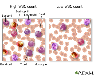
This picture above shows the different kinds of white blood cells. WBC'S are categorized into groups. The granular leukocytes, which have a granular appearance and the Agranular leukocytes which do not have a granular appearance. There are 3 types of granular leukocytes.
1. neutrophils - which are the most abundant of WBC's. Usually first responders to invasion by a pathogen.
2. Eosinophils - increase in number in parasitic worm infection
3. Basophils - similar to mast cells. They release histamine when allergic reactions happen.
The other group of WBC's, the Agranular leukocytes have just two types;
1. Lymphocytes - responsible for immunity to particular pathogens and their toxins. There are 2 types of lymphocytes: a. B cells - these are responsible for specific immunity by producing antibodies
b. T cells - these are also responsible for specific immunity by producing antibodies, but one T cell, the cytotoxic T cell directly destroys pathogens.
DISORDERS WITH WBC'S
Severe combined immunodeficiency disease is a WBC disorder. I actually remember the movie "The boy who lived in a Bubble." This disorder is when the the stem cells of WBC's lack an enzyme called adenosine deaminase. People without this enzyme cannot fight off infections. The boy in bubble was protected from the outside world. He could be compromised while in that bubble. Unfortunately, he died when the doctors tried injecting his bone marrow with donated blood cells. Another disorder is leukemia, this is a form of cancer and WBC proliferation. The WBC's are abnormal or immature.
BLOOD CLOTTING AND DISORDERS
Blood clotting is very important when it comes to an injury (small one) because when cut the blood rushes to the injury and forms a clot. When the injury first happens platelets and damaged tissue release prothrombin activator and converts the plasma protein prothrombin to thrombin. Thrombin acts like an enzyme the severs two short amino acid chains form each fibrinogen molecule. These activated fragment join to form long threads of fibrin. which covers the blood and forming a clot so blood will not escape anymore.

This is what it actually looks like. Some people have clotting disorders. Thrombocytopenia is an insufficient number of platelets. If your blood does not clot and you have injury to your body, you could bleed to death because the clot will not form at site. Another disorder is hemophilia. Hemophilia is a rare, inherited bleeding disorder in which your blood doesn’t clot normally. If you have hemophilia, you may bleed for a longer time than others after an injury. You also may bleed internally, especially in your knees, ankles, and elbows. This bleeding can damage your organs or tissues and, sometimes, be fatal.
ABO BLOOD GROUPS

This picture is great example of how to find out your blood type, only if you had the antigens to mix with your blood. We did this in my physiology class. As you can see, the blood cells and the antigens that are compatible with it. I think this picture really shows what I would of tried to explain. It is important to know your blood type if you ever need a transfusion. The blood types have to be them same in order to get the transfusion. Another blood type the Rh- and Rh+. usually occurs within a pregnant women.

In this pregnancy the child is Rh+ and the mother is Rh-. The Rh negative mother can start to produce antibodies against the Rh+. With another pregnancy and another Rh+ baby and the anti Rh antibodies may cross the placenta and destroy the child's RBC's. This is called hemolytic disease. If the RBC's are destroyed there is hemoglobin breakdown products in the blood can lead to brain damage and mental retardation, and sometimes death.
HOMEOSTASIS
Homeostasis is the maintenance by the highly coordinated, regulated actions of the body systems of stable chemical and physical conditions in the internal fluid environment that bathes the body's cells. Without homeostasis we would not survive. Homeostasis makes everything run smoothly as long as all systems are not compromised.
LYMPHATIC SYSTEM AND IMMUNITY
a. microbes, pathogens & you
b. bacteria
c. viruses, prions
d. Lymph system and organs
e. inflammatory response
f. defenses ( t cells and B cells)
g. Active & passive immunity
h. disorders of the immune system
Microbes and pathogens can be found everywhere and anywhere. We even carry some, mostly non-pathogens. They can be spread about in numerous ways. If someone has TB (tuberculosis), and they sneeze, if you are within 3 feet of them you have a very good chance of getting it. Our bodies have lines of defense. One being our skin, this is the first line of defense. Secondly, is the mucous membranes of our body cavities. An example of a mucous membrane would be the the respiratory system, which is lined with epithelium tissue.
BACTERIA
Bacteria are prokaryotic and they lack a nucleus. They come in three shapes, bacillus (rods), coccus (spherical), and spirillum (spiral).

Bacteria have a cell wall made of peptidoglycan. They reproduce by binary fission which is they produce two cell that are identical to the original cell. Some cell walls are surrounded by a capsule that has a thick gummy consistency. Bacteria also have appendages that help them move about. They are called flagella and some have fimbriae which are stiff fibers that allow bacteria to adhere to surfaces like "host cells." Another appendage is a pilus, this is a elongated hollow appendage used to transfer DNA from one cell to another.
VIRUSES
Viruses can enter the body through any opening. I don't think I have to point those out on our body, but this is how they enter. Viruses are acellular and do not live independently. Some viruses that many are familiar with is AIDS, chickenpox, and the flu. A virus has two part the outer capsid composed of protein unites and an inner core of nucleic acids. Viruses carry their own genetic code so that they can reproduce by itself. It just uses our bodies as a "Host." Some viruses are being transported from other countries. Our nation is worried about a pandemic! One example of this is the bird flu which started in Asia and was transported to Canada.
PRIONS
Prions are proteinaceous infectious particles, what this means is that a regular protein is compromised and cannot perform its regular functions. Prions cause degenerative diseases of the nervous system. One very familiar prion is the Mad cow disease. This disease was thought to have been transmitted by ingestion of brain and nerve tissue from infected animals.
LYMPHATIC SYSTEM
This system consists of lymphatic vessels and organs. The function of the lymph system is the return of excess filtered fluid. Lymphatic vessels form an extensive and complex interconnected network of channels. They mainly collect fluid lost from vascular capillary beds during nutrient exchange and deliver it back to the venous side of the vascular system. The lymphatic system as two ducts; the thoracic duct which returns lymph collected form the body below the thorax, the left arm, and left side of the head and neck into the subclavian vein. The second duct is the right lymphatic duct this duct returns lymph form the right arm and right side of the head and neck into the right subclavian vein. The lymph organs consists of the red bone marrow, and the thymus gland and there are secondary organs which are the lymph nodes and spleen. Red bone marrow produces all the the blood cells.

The thymus gland, (seen in this picture) has two functions. It produces thymic hormones, such as thymosin which are believed to help in the maturation of T cells. Secondly, immature T cells travel to the thymus gland where they can mature. The lymph nodes are found all over our bodies, for example if you feel a lump under your arm that is a swollen lymph node. The lymph nodes are divided into compartments. The compartments each contain a sinus that increases in toward the center of the node, therefore making the node swell. The spleen is also filters blood and plays a role in the lymph system. The spleen is also considered the largest organ of the lymph system. The spleen is also divided into sections in called the red pulp and the other white pulp. The red pulp surrounds venous sinuses is involved in filtering blood. The blood must pass through the sinuses before exiting. Here the blood is cleaned of debris.
INFLAMMATORY RESPONSES
Inflammatory responses are our bodies protected where the inflammation is taking place. This mechanism recruits the WBC's to help in the defense against infection. Chemical mediators also help with the defense because they cause the capillaries to dilate and become more permeable. The site of injury can turn red due to the excess blood flow through the capillaries. Increase in body temperature can prevent the spread of pathogens because they can't grow and increased blood flow brings more fighting WBC's. If the injury is not severe the redness and temp will go away shortly, but if the injury is severe the neutrophils (wbc), can secrete chemical mediators called cytokines. These attract more WBC to the area of the injury. Monocytes are also attracted to the area. Monocytes become macrophages and are more helpful the monocytes because they can get lymphocytes to carry out defense mechanisms.
DEFENSE WITH T CELLS AND B CELLS
These two lymphocytes are very important in helping our body with defense!! We have antibodies in our bodies that fight off antigens, but we need more. When the antigen is more powerful, then we need the help of these lymphocytes. They both have receptor that aid in recognizing antigens. The B cells produce plasma cells and memory cells. The plasma cells circulate in the blood and lymph. They are very large for the mass production and secretion of antibodies to a specific antigen. The memory cells produced by the B cells are just what they are called because if the same antigen entered the body the memory cell divide and give rise to more plasma cells capable of producing the correct type antibody.
ACTIVE & PASSIVE IMMUNITY
Active immunity can involve a pathogen that you have had and your body builds an immunity to it, meaning you will not get it again. People can also be immunized from getting a disease. The person is injected with with the pathogen or a weaken version of the pathogen and the body build antibodies. These immunizations can not cause the pathogen to become active because they are no longer virulent. After you have been immunized against something specific you can see if the antibodies have built up by having a blood test called a titer. This blood work will show if you have a immunity to that specific pathogen. Passive immunity is when a person is given prepared antibodies or immune cells to combat a disease. This type of immunity does not last. One example is the newborn being exposed to something but the mothers antibodies have passed through the placenta protected the newborn for a short period of time. Also, if the mother breast feeds, her antibodies pass through the milk and protects the infant .
DISORDERS OF THE IMMUNE SYSTEM
The disorders of the immune system involves autoimmune disease, which causes the body to attack itself. The is unknown causes why these diseases occur. One disorder is Multiple Sclerosis, in which the T cells attack the myelin sheath of the nerve fibers and causes neuromuscular symptoms like, leg weakness leading to the inability to walk. Another autoimmune disease is Rheumatoid arthritis where the joints are affected and the joints affected look deformed.

When a persons immune defense is compromised they are unable to protect themselves from disease. AIDS is another example of a autoimmune disease, or acquired immune deficiency. People with AIDS are a very weak immune system and are susceptible to infections. The can contract pneumonia, cancer, and other viruses.


No comments:
Post a Comment