Wednesday, October 29, 2008

COMPENDIUM III THE NERVOUS SYSTEM

 www.completehealings.com


Nervous system

a.  nervous tissue and functions

b.  myelin sheath

c.  nerve impulses and propagation of action potentials

d.  sodium/potassium pump

e.  synapse

f. CNS

g.  PNS

h. Effects of substance abuse

SENSES

a.  receptors and sensation

b.  proprioceptors, cutaneous, and pain receptors

c.  sense of taste and smell

d.  sense of vision, A & P of vision

e.  sense of hearing, A & P of hearing

The Nervous system contains two major regulatory system of the body.  There is the Central Nervous System (CNS), and the Peripheral Nervous System(PNS).  First I would like to explain nervous tissue and their functions because this is how these systems function properly. 

Nervous tissue contains two types of cells; neurons and neuralgia (cells).  The neuron plays a significant roll in the Nervous system.    There are three parts to a neuron.  First there is the cell body which contains the nucleus, mitochondria and other organelles.  Next, there is the axon, which conduct impulses away from the cell body.  Dendrites are the last part of a neuron.  They are responsible for receiving messages from other cells and conducting those impulses towards the cell body.  

www.understanding_ocd.tripod.com

There are also three types of neurons and they are classified by their function.  Sensory neurons, these neurons are responsible for carrying impulses from the sensory receptors to the Central nervous system.  Motor neurons, transmits the impulses or messages from the CNS to a effector muscle or a gland.  Effectors carry out our responses to environmental changes, being internal or external.  Interneurons, they are only found in the CNS and connect neuron to neuron.  They also communicate the messages to the motor neuron, but before they do they sum up the information.  Sensory receptors are special structures that detects change in the environment.  Neuralgia cells (glia cells) cannot conduct their own action potentials and serve as support cells and help protect neurons.

Myelin Sheath is the protective covering surrounding some axons.  The myelin sheath is composed of schwann cells, which are produced in the PNS.  The cells wrap themselves around the axon several times.  In the CNS, oligodendrocytes are responsible for the myelin sheath.  Both of these cells only cover a portion of the axon, leaving exposed areas, which are called Node of Ranvier.  Gray matter in the CNS is gray because it does not contain myelinated axons and the white matter in the CNS is white because it does contain myelinated axons.  Important note is that if an axon is accidentally severed the myelin sheath remains and serves as a passageway for new fiber growth.

ACTION POTENTIAL

This is a great example of an action potential.  I will explain each of these processes that appear in this graph I found online.  Action potentials are measured my an voltmeter.  Action potentials are the rapid change in polarity across an axonal membrane as the nerve impulse occurs.  All of these steps ARE the action potential.   I will try and go through each step and how they occur.  Resting potential is when the axon is not conducting an impulse or a message.  The resting potential usual measures at -65mV (millivolts).  When the membrane is stimulated it creates an action potential. POLARIZATION, not seen in this graph charges are separated across the plasma membrane has potential.  Anytime the value of the membrane potential is other than )mV in either positive or negative direction the membrane is in the state of polarization.   When DEPOLARIZATION occurs a change its the potential that makes the membrane less polarized (less negative,inside) than at resting potential.  Depolarization decreases membrane potential moving it closer to 0mV.  Notice the graph above.  With REPOLARIZATION,  the membrane returns to resting potential after having been depolarized.  HYPERPOLARIZATION,  is a change in potential that makes the membrane more polarized (more negative) then at resting potential.  It increases membrane potential moving it even farther from 0mV.   All of these actions are controlled by voltage gates and the sodium potassium pump.    These gates open and close allowing ions (sodium and potassium) into and out of the cell.  The sodium gates open first when an action potential occurs and Na+ (sodium) flows into the axon.  When it moves inside it changes the membrane potential from negative to positive.  The potassium (K+) gates second and the potassium flows out changing the membrane potential.  The sodium-potassium pump is very important when it comes to balance.  Both of these ions are very important in  homeostasis!!!!  

www.upload.wikimedia.org/wikipedia/commons

Propagation of an action potential.   I found this great animation online showing the action potential.  When the axon is unmyelinated the action potential at one location stimulates an adjacent part of the axons membrane to produce a action potential.  In myelinated axon there are bare spots on the axon itself (node of Ranvier) that speeds up the action potential.  This is called a saltatory conduction.  The nerve impulse actually jumps from node to node.  After a stimulus of an action potential occurs, it goes through a refractory period.  This is when the patch of membrane that has just been stimulated it becomes unresponsive to further stimulus and prevents an action potential from spreading backwards into the area through which it just passed.  An action potential is an all-or-none event!!!

SYNAPSES

A synapse is actually a junction between a nerve cell and another cell.  Action potentials are propagated from the axon hillock to the axon terminal.  The axon hillock is the first portion of the axon where the action potential takes place then it makes its way down the axon to the axon terminals.  At the synapse, there is a small gap called the synaptic cleft, this area is actually to wide for the direct spread of a current from one cell to another.  It prevents action potential from electrically passing between neurons.   Synapse only work in one direction.  Neurotransmitters carry the signal across the synapse.  The neurotransmitters are stored in the axon terminals. 

www.content.answers.com/

STEPS IN THE SYNAPSE

1.  When an action potential in a presynaptic neuron has been propagated to the axon terminal, this change in potential triggers the opening of the Ca+ (calcium) gate  channel in the synaptic knob.

2.  The calcium ions flow through the opened channels

3.  Calcium allows the release of a neurotransmitter from some of the synaptic vesicles into the synaptic cleft.

4.  The released neurotransmitters diffuse across the cleft and binds with protein receptor sites.

5.  The binding triggers the opening of specific ion channels, changing the ion permeability of the postsynaptic neuron. 

6.  Once the neurotransmitter has been released and initiated a response, it is removed from the cleft.

Some synapses can be excitatory or inhibitory.  With the excitatory synapse the response to the binding of a neurotransmitter to the receptor is the opening of nonspecific channels and permit passage of sodium.  With inhibitory synapse it would allow the passage of potassium to enter the neuron.  Sometime a neurotransmitter can be inactivated due to an enzyme called acetylcholinesterase.  This particular enzyme breaks down acetylcholine.  Some neurotransmitters are reabsorbed for reuse.   There are different types of neurotransmitter molecules.  One example, is acetylcholine and norepinephrine.  These two are seen in the CNS and the PNS. Norepinephrine excites smooth muscle.

CENTRAL NERVOUS SYSTEM

www.medem.com/MEDEM/images


The central Nervous System consists of the spinal cord and brain.  The brain is where the sensory input is received and then sent to the motor control for initiating.  The brain and spinal cord are protected, not only by bone, but other important membranes.  Underneath the bone are the meninges and the spaces between these meninges is cerebral spinal fluid which cushions and protects the organs they hold.  There are three layers to the meninges.  The first layer is the PIA MATER (delicate mother) and this layer is vascular and shares blood supply with the underlying nerve tissue.  The second layer is the ARACHNOID MATER and this protects the brain.  It also resembles a spider web, contains cerebrospinal fluid and is impermeable.  The third layer is the DURA MATER (tough mother) and is functionally important because it reduces the risk of abrasion of the inner layer and the central nervous system.  The brain also has four ventricles which are interconnecting chambers that produce and serve as a reservoir for cerebrospinal fluid (CSF).  This reservoir is very important because it drains the excess CSF into the cardiovascular system.  If it is not drained, with a child it can cause hydrocephalus which is water on the brain and in an adult the extra fluid has no where to go because the adult brain is larger then a child's that is still developing.    The CNS is composed of two types of nervous tissue; GRAY MATTER and WHITE MATTER.  The Gray matter is mostly cell bodies,dendrites and terminals, is the spinal reflex integrating center and consists of sensory and motor nuclei.  The White matter have bundles of myelianted axons and run along tracts.  These are ascending tracts with sensory to dorsal (the back) root that carry info to the brain.  The descending tracts are motor to ventral (the front) roots and carry commands to motor neurons.  The spinal cord is on the dorsal side of the body and extends from the base of the brain through the foramen magnum and the vertebral column.  The bones in the vertebra protect the spinal cord.  The spinal cord plays a key role in communication between the brain and the peripheral nerves. 

www.serious-injury-lawyers.org

The spinal cord is the location where reflex arc happens.  An example of a reflex arc would be a painful stimulus.  When this happens it sends a signal to the brain through the afferent pathway, which then propagates the electrical signal.  Then once the afferent neuron enters the spinal cord it diverges or spreads and terminates on three different types of internuerons; (1) excitatory interneurons which will in turn stimulate the efferent motor neurons to the site where the pain was felt and pull it away from the stimulus (2) inhibitory interneurons will inhibit the efferent motor neuron to the site of the pain and preventing counterproductive contraction of this muscle and (3) interneurons  that carry the signal up the spinal cord via the ascending pathway to the brain for awareness of pain and memory storage.  Before I move on I would like to point out some portions of the brain.  The cerebrum, (telencephalon) is the largest portion of the brain and is located in the frontal lobe, which is the forehead area.  The cerebrum is the last area that receives sensory input.  The cerebrum is responsible for reasoning and thought.  It has two parts to it and is called the cerebral hemisphere.  It is further divided into left and right.  Each of these hemispheres are divided into four lobes; frontal,parietal, temporal, and occipital.  The cerebral cortex plays a key role in the most sphicated neural functions such as, voluntary initiation of movement, final sensory perception, conscious thought, language and personality traits.    The cortex contains both sensory and motor areas.    Another interesting fact about the cortex is where language is associated.  the Wernicke's area and Broca's area.  The Wernicke's area helps us understand written and spoken language and then sends info to the Broca's area.  The Broca's area  stimulates the right muscles for speaking and writing.  The hypothalamus and thalamus are located in the dienephalon, which is in the  third ventricle.    The hypothalamus is associated with the pituitary gland and controls body temp, urine output, uterine contractions and is the thalamus receive sensory input except smell.  The cerebellum lies in the occipital lobe (vision) and is actually separated from the brain stem by the fourth ventricle.  The cerebellum is responsible for coordination of muscles, balance, posture and muscle tone.   The medulla oblongata is very important because it is responsible for heart beat, breathing, coughing, sneezing, and blood pressure.

LIMBIC SYSTEM

The Limbic system is involved with our emotions and motivations.particularly those that are related to survival. Such emotions include fear, anger, and emotions related to sexual behavior. The limbic system is also involved in feelings of pleasure that are related to our survival, such as those experienced from eating and sex.  There are two significant structures in the Limbic System they are the amygdala and the hippocampus.  The amygdala is involved in emotional responses, hormonal secretions, and memory.  It can use past knowledge to assess a current situation.  The hippocampus is responsible for sending memories out to the appropriate part of the cerebral hemisphere for long-term storage and retrieving them when necessary.  Also, plays a key role in learning and memory.  There are different types of memory; short term, long term, semantic, and episodic. 

PERIPHERAL NERVOUS SYSTEM

The PNS contains the nerves which lie in the spinal cord.  We have 12 pairs of cranial nerves.  They all serve a different purpose; mainly the head, neck and facial region.  We also have 31 pairs of spinal nerves.  The PNS consists of nerves that transmit info to the and from the CNS.  Sensory(afferent) neurons transmit nerve impulses towards the CNS.  Motor (efferent) neurons transmit impulses away from the CNS towards the effector organs such as muscles or glands.  The PNS also has divisions;  The autonomic which divides further into the sympathetic and parasympathetic and then the somatic system.  The nerves in the somatic system serve the skin, skeletal muscles and tendons.  The autonomic systems with its two divisions are interesting.    Here is a great picture separating the the two systems and their actions to stimulus:

www.biocomtech.com

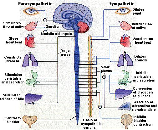
As you can see in the picture above these two divisions of the autonomic system have opposite effects to certain forms of stimulus.  The parasympathetic slows things down, and the sympathetic speeds things up.  Also, in this picture you can see the nerve that innervates these reactions and parts of the brain where it is happening.

EFFECTS OF SUBSTANCE ABUSE

When you think of substance abuse some just think of heroin or pot, but there are so many (more today) "substances" out there that can effect your brain function and  even damage brain cells.  Alcohol is a drug that is accepted worldwide.  There are a lot of people who abuse alcohol and is known to have harmful effects on the body and the brain.  Not only does it effect the brain, but it can damage the liver and lead to death.  The liver acts as a filter and gets rid of the bad stuff, but drinking alcohol excessively can damage that part that is so important.  Alcohol also impairs our thinking, reasoning, and coordination.  Alcohol is a depressant and slows down your respirations which can lead to death.  some people can consume to much alcohol (effecting the CNS) and get alcohol poisoning.   Another drug that people may not even consider a drug is nicotine.  Nicotine has effect on the CNS and the PNS.  With the CNS it binds to neurons within the CNS and dopamine is released.  Dopamine will increase your heart rate and blood pressure.  This constant effect will damage your heart.   Another drug is methamphetamine.  This is extremely dangerous because some people create meth labs which contain explosive stuff.  Meth, as it is called, when smoked can keep the person awake for days.  This obviously can be detrimental to the human body.  There are a lot of drugs in the world today that can be deadly.  The list can go on forever.  I am sure I haven't heard all of them. 

SENSES

RECEPTORS AND SENSATIONS

There are different types of receptors.  First there is sensory receptors and these are familiar because of the nervous system.  The sensory receptors are dendrites that detect certain types of stimuli.  Exteroceptors are ones that detect stimuli form the outside of the body, such as taste and smell.  Interoceptors receive stimuli from the inside of our bodies.  Examples of interoceptors are pressorecptors which respond to changes in blood pressure.  Osmoreceptors detect change in the water-salt balance and chemoreceptors monitor the pH of the blood.  Interoceptors are directly involved with homeostasis!  

TYPES OF SENSORY RECEPTORS

Chemoreceptors have to do with chemicals.  As you can see by there name; "chemo"  They are sensitive to specific chemicals, they include the receptors for smell and taste and as well as those located deeper within the body that detect oxygen and carbon dioxide concentrations in the blood or the chemical content in the GI system

Pain receptors (nociceptors) are sensitive to tissue damage such as pinching or burning.  They make us aware of possible danger.

Photoreceptors are responsible for vision.  The visible wavelengths of light.  The photoreceptors can be found in our eyes.

Mechanoreceptors are sensitive to mechanical energy.  One example is the skeletal muscle is sensitive to stretching or the ear containing fine hair cells for hearing are bent due to sound waves.

Thermoreceptors are sensitive to heat and cold.

Sensations involve the sensory receptors because it is a response to the sensation that makes the receptors to respond.   When the nerve impulses arrive at the cerebral cortex of the brain, sensation which is the conscious perception of stimuli occurs.  Above I explained the reflex arc and this is exactly like that.  The great example of sensing pain which in turn will alert the response of pulling away from it.

PROPRIOCEPTORS, CUTANEOUS RECEPTORS, PAIN RECEPTORS

Proprioceptors are responsible for knowing the position of our limbs in space by detecting the degree of muscle relaxation, stretch and movement.   The cutaneous receptors are in our skin.  It is in the deep within the dermis layer.  These receptors are responsible for alerting us of touch, pressure, pain, or temperature.  Pain receptors alert us of pain, whether it be internal or external. 

SENSES OF TASTE AND SMELL

Taste and smell are chemoreceptors because they are sensitive to foods we eat and the air we breathe.  Sense of taste is due to the "taste buds" on our tongue.  The tongue is divided up making certain areas responsible for sweet, sour, salty, and bitter.  These four are common, but there is another called umami which may exist for certain flavors such as cheese, beef broth and some seafood.  The gustatory cortex is responsible for the interpretation of particular tastes. The olfactory cells in our noses is responsible for the sense of smell and what we perceive as taste is due to smell.  There are different kinds of odor molecules and attracts a combination of receptor proteins.  Stimulates different combination with different odors.

SENSES OF VISION, A & P

www.eyesareus.org/web/eyesareus.org

In order for us to see it requires the help of the brain.  There is a optic nerve which sends a message to the brain.  This picture above shows the human eye and all the things that are involved.  I will explain each of the components of the eye.  There are three specialized tissue layers.  The sclera, which is the white part of the eye along with the cornea (is not white), the choroid/ciliary body,  iris are the middle layer underneath the slcera; the choiroid layer becomes specialized to form the ciliary body and the iris, and the retina which is the innermost layer.   Between the lens and the retina there is a semi-fluid the vitreous humor and is important in maintaining the spherical shape of the eyeball.  Between the cornea and lens contains a clear watery fluid the Aqueous humor and this carries nutrients for the cornea and lens both of which lack blood supply.  Also the aqueous humor is produced by a capillary network within the ciliary body and if it is not drained it could block drainage canals.  The round opening in the center of the iris is the pupil, which constricts or dilates to adjust to light.  The iris is responsible for controlling the amount of light entering the eye and regulates the size of the pupil.  Sensory tunic (retina) contain rod and cones.  Rods make you see black and white and cones provide colors.  Signals pass from photoreceptors and leave the retina toward the brain through the optic nerve transduction.   The fovea centralis is the area of the retina with only cones.  There are three types of cones that detect different colors (green,red,blue) and if you lack one type it causes colorblindness.  The blind spot is located at the optic disc.   When trying to focus (when looking at something), there are two things that are happening with the cornea and the lens.  Rays from light more then 20ft away are considered parallel by the time they reach the eye.  From near objects the light is still diverging (radiating outward) when they reach the eye.   Accommodation increases the strength of the lens for near vision.  The function of the photoreceptors is the rod and cones. The rods contain a pigment which is purple called, rhodopsin.  Rhodopsin is a molecule made up of protein opsin and a light absorbing molecule called retinal.  When a rod absorbs light rhdopsin splits into opsin and retinal.  Rods are suited to night vision.   The retina contains ganglion and bipolar cells.  The ganglion cell axons go to the optic nerve.  Start of an action potential cones out the way light came in.  Light enters the retina on the side containing the optic nerves and travels through all the layer before reaching the receptors.  The optic nerve lacks receptors.  A visual pathway to the cerebral cortex------>begins at the retina------>light activates photoreceptors----->photoreceptors signal bipolar cell-----> bipolar cell signal ganglion cell--------> axons of ganglion cell exit the eye as the optic nerve.  There are disorders with the eyes. 

SENSES OF HEARING, A & P

https://www.lpch.org/photos

The ear is not only for hearing, but it involves our balance (equilibrium) too.  This is a picture of the outside of the ear as well as the inside.  I will start with the outer ear.  The outer  is composed of the auricle  (earlobe) and that helps direct sound.  External auditory meatus (entrance to inner ear) is lined with skin, contains hairs, sebeous glands and ceruminous fluid (earwax).  The tympanic membrane forms the boundary between the external and middle ear.   The middle ear consists of the tympanic cavity a small air filled space located within the temporal bone.  Medial wall contains oval window membrane where the stapes (bone) is attached.  The round window another small membrane covering the opening seals the lower compartment from the middle ear.  Pharyngotympanic tube or Eustachian tube links the middle ear to the pharynx.  These are normally collapsed but if you yawn they will open.  The ear ossicles are in the middle ear and they are the stapes (vibrates against the oval window), malleus (attach to eardrum), and the incus (between the malleus and stapes.  The inner ear also called the labyrinth (maze) lies within the temporal bone.  bony labyrinth is a cavity consisting of 3 parts; (1) the semicircular canals which functions in orientation eg., gravity), (2) vestibule conducts sound waves (3) cochlea-contains receptor hairs, also the hearing portion of the ear.  The membranous labyrinth fits within the bony labyrinth and also has 3 parts; (1) semicircular ducts-ampulla houses a structure call a crista ampullaris and these contain receptor cell of rotational acceleration, (2) utricle and saccule these are suspended in the perilymph and are otolith organs (provide info about the position of the head), (3) cochlea duct the middle ear compartment tunnels through the center of the cochlea. The organ of Corti is responsible for hearing.   The process of hearing starts with sound waves that travel.  hearing reception------>sound vibration----->tectorial membrane movement---->bends sterocillia--->stimulates electrical response-------->fires synapses-------->auditory axons carry signals.   If your tectorial membrane is damaged you will lose your hearing.  The vestibular nerve helps us maintain our equilibrium.    All of our senses are very delicate.  With having a lot of "tiny parts", if one tiny piece is missing then those senses don't work properly. 

Wednesday, October 15, 2008

SELF EVALUATION UNIT II


The things about Unit II that I am most proud of was my lab project until I posted it.  It looked great before I uploaded it!!!  I am also happy that I completed all the assignments.  That is always a great accomplishment when things are done!!  The last thing I am proud of is that  you can actually see all of my pictures and don't have to put your nose to the screen.

Would I make any improvements??  Umm, sure there is always room for improvement.  I will always say that I have a hard time putting things into words, but I do my best.  I am still trying to get use to the "blog" thing and I will probably have it mastered right before this class ends!!

My overall grade should reflect the quality of my work.  I believe with completing the assignments shows that I care about the dead line and want it to be right!  I think I put a lot of effort into any work I do, so I guess I feel deserving of a good grade.

In the next unit I could do better by reading the lab project thoroughly before I start it.  I didn't think that this one was going to be a lot of work, but it was.  The one on the first unit was too, but being in the medical field I saw that we had to do blood pressure and stuff like that so I wasn't really concerned. 

I felt most engaged with the circulatory system.  I think that it is so fascinating!!  Actually all of the chapters were great.  Genetics is not my thing but getting into the systems in the body is what I enjoy the most.

I don't think I ever felt distanced from the course because I am busy trying to get the work done.  I get nervous knowing the due date is around the corner.

What I found helpful in this particular unit was all the extra web sites that the instructor made available.  It is always helpful to have more information.

I didn't find anything puzzling.  I think Unit II was a great refresher for me!

The thing that surprised me about this Unit was doing the lab exercise.  I have been working out since I was in my teens and when I saw some of the blood pressure and pulses readings it made me a little nervous.  My blood pressure has always been great.  When I did some of the exercises and then took my blood pressure and pulse, I was surprised how high they were.  I know when we did stress test in the doctors office I worked in the patient had to reach a certain target rate(pulse) for their age.  I think during my lab project my pulse was a little too high.

EXERCISE PHYSIOLOGY PROJECT UNIT II

WWW.howstuffworks.com/gif/adam/images/en/1

This lab project is to see how my blood pressure, pulse, and respirations are affected  by doing physical activities.  I chose physical activities because I try to exercise at least four times a week.

I have chosen running around my house for the first exercise assignment.  I am literally running outside around my house.  For this exercise, I will run around my house once, then come in and take my measurements.  I know that my metabolic measurements will go up because this exercise is physical.  When you exercise, your heart rate goes up, so with my heart rate up, that makes the heart beat faster, and forcing blood through faster.   The blood moving faster through my heart will make my blood pressure go up.  The heart has to work harder then at rest.  With the repetition of running around my house, (resting in between, through the course of the day) I believe that my measurement will go up, but that they may stay the same with each repetition.  While resting, my measurements should come back to, or close to my baseline, I am not sure how long this takes, but I guess about a half hour.

For the second exercise, I chose to lift weights.  This is a tuff one, but I believe that my metabolic measurements will go up slightly.  With the repetition of doing this three times through the course of the day I will increase the number of time I lift them.  For the first repetition I will do four sets of  6 reps (arms), for the second, I will do six sets of 6 reps, and for the last one I will do eight sets of 6 reps.  I know that my measurements will go up, but it won't be as drastic as running around my house.  After I rest, I believe that my measurements will return to my baseline.  Lifting weights is physical, but not as physical as running.  My recover for my measurements should take about a half hour.

For the third exercise, I have a manual treadmill that I will walk on.  First for 30 seconds (not easy to move)!!!  Second, I will do one minute and the third I will do a minute and a half.  My treadmill is not electric so it is very hard to move so I know that my measurements will go up.  The one for 30 seconds will not  affect my measurements to much, but the next two will.  I will also rest in between these reps.  My measurements should go back to baseline.  I am not sure how quickly your measurements recover for activity, but I believe it should take about a half hour to 45 minutes?!

Pics

devices used This is a picture of my digital blood pressure machine and a watch to take my pulse and respirations.

takingbp This is me taking my blood pressure

digitalbp The reading of my systolic and diastolic.  This machine also takes my pulses, but I used my fingers and a watch.

takingpulse This is me taking my pulse

treadmill This is my manual treadmill and on it is  the sneakers I used to run around my house and the weights that I used for my 2nd activity.

As for my hypothesis I think I was very close to what I imagined.  With my first exercise, running, my measurements went up drastically.  Kind of scared me!  My second activity, weights, I  said that they would go up slightly and they did.  The respirations went up the most.  As for the third activity, the treadmill, all the measurements went up.  The first reading only went up slightly because I was only on the treadmill for 30 seconds.

Here are my graphs.........

Preview of your graph

Below is a preview of your graph. To edit your graph Click here...

Preview of your graph


Preview of your graph


Below is a preview of your graph. To edit your graph Click here...

Preview of your graph

I tried to put the metabolic table that you provided, but it would not cut and paste.  So I tried my best.


METABOLIC RATE DATA TABLE

Repetition 1    Rep 2    Rep 3    Rep 4    Rep 5                                            Means
Baseline    resting    resting    resting    resting    resting   
PULSE
68    66    66    64    66    70                                                                             66.4

RESPIRATION
20    18    18    20    18    18                                                                              18.4
 SYSTOLIC
108    106    111    111    102    96                                                                    105.2
DIASTOLIC
78    65    70    72    65    59                                                                               66.2
Activity 1    Running                    
PULSE
68    119    88    101                                                                                          102.6
RESPIRATIONS
29    26    24    26                                                                                                25.3
SYSTOLIC
108    137    151    149                                                                                       145.6
 DIASTOLIC
78    88    91    104                                                                                              94.3
Activity 2    Weights 4 sets    6 sets    8 sets           
PULSE
68    86    80    84                                                                                                 83.3
RESPIRATIONS
20    18    28    28                                                                                                 24.6
SYSTOLIC
108    115    112    107                                                                                         111.3
 DIASTOLIC
78    76    70    63                                                                                                  69.9
Activity 3    Treadmill 30sec    1 min    1.5 min       
PULSE
68    93    127    125                                                                                                115
RESPIRATIONS
20    22    28    30                                                                                                    26.6
SYSTOLIC
108    108    128    135                                                                                            123.6
DIASTOLIC
78    78    82    85                                                                                                     81.6

ETHICAL ISSUE UNIT II WHAT IS FOOD?!


The picture above represents what all the doctors, the media,the FDA, etc. say we should eat!  This pyramid was created by the FDA to let people know the food groups that are important in our diets.  So why don't people follow this so called guideline?!!  Well, what about single mothers or fathers struggling to make ends meet and putting healthy food on the table.  Not an easy task!!   The only thing they can afford is the processed food, packaged foods, and what ever else is on sale.  Things like, Kraft macaroni and cheese on sale for......6 boxes for a 1.00 or hamburger helper two boxes for 5.00.  These two examples are horrible for you.  Sure people could buy produce for salads, and a bunch of fruit, but their dollar stretches a lot further with the foods I just mentioned.  Healthy food is more expensive!!  Isn't is sad "they" say eat healthy, but yet a lot people can't afford to do that.  They wonder why obesity is on the rise in Americans, especially in children.  Type II diabetes is becoming common in children and that is scary to me!!  The more expensive foods I am talking about is organic foods because they are the safest and don't contain any chemicals. Understandably, it "may" cost more to produce organic foods, but if they sold it at a reasonable price they may sell a lot more because  people could afford it.

I get so sick and tired of hearing what the media has to say about what you should and should not be eating, what is healthy, what causes cancer, and on and on.  I am sure a lot of people would agree with me.  The stress of trying to eat right is frustrating.    

I grew up with a single parent.  My mother worked three jobs while supporting six children.  As you can imagine, we ate a lot of  macaroni and cheese and a lot of stuff out of a can because my mother wasn't home to cook that much.  So as I was growing up I was a chunky kid, but again as being a teen, peer pressure and the media once again step in and remind you to eat right, stay slim.  Nutrition is very important for children because it helps them grow probably.  Children that suffer from malnutrition have stunted growth, abnormal abdomens, and are very weak!  How about school lunches?   You have probably seen in the news that some schools are trying to change the menus and make them more healthy, but for now most of the lunches have fried foods, hamburgers, or pizza.  I am not saying having these are bad for children, what I am saying is moderation is the key!!!    Anything in access is not good for you.

I really enjoyed the "farm to school online.  What a great idea!!!  Not only will the children be eating right, they can learn how to grow their own stuff  at home.  How rewarding is that!!!  I eventually, because of my older sister, started growing my own vegetables, herbs, and flowers.  There is nothing like bringing your produce to your table!!  Since moving to Arizona from RI I have not had the chance to grow anything because I have only been here for 4 months, but I have visited a lot of the nurseries!  It really is amazing what you can do with ONE seed!

As for thinking of ways to solve obesity or nutrition problems, I really think it comes down to affordability.  Some people are raised with poor nutrition, but the media is always there to remind you what you should be eating and lets not forget exercise.  I think people have to start making healthier choices too. 

Monday, October 13, 2008

COMPENDIUM UNIT II

            DIGESTIVE SYSTEM & NUTIRTION


WWW.apps.uwhealth.org

DIGESTIVE SYSTEM

a.  Overview

b.  wall of the digestive tract

c.  where digestion begins and ends

d.  complications with poor nutrition

e.  disorders of the liver, colon

NUTRIRION

a.  weight control, obesity

b.  nutrients and vitamins

c.  food guide pyramid

d.  eating disorders

I think the digestive system is one of the more interesting system in our bodies.    The digestive system consists of many organs working together.  The digestive system is also referred to as the GI tract (gastrointestinal tract).  It starts with the mouth because this is where ingestion begins.  Our mouths contain saliva and enzymes that help, along with our teeth, break down food and turn it into a bolus.  The bolus is a soft ball of broken down food that can be easily swallowed.   This might also be considered digestion because of the breakdown of food. All parts of the digestive tract contains digestive enzymes that hydrolyze foods to molecular nutrients for our bodies to absorb.  The GI tract has three main functions; movement, this is due to peristalsis.  Peristalsis is involuntary muscle at work.  Food must has to pass along form organ to organ and peristalsis makes that happen.  Another function of the GI tract is absorption, this happens when the food has been broken down into molecules and can be absorbed by the body.  The third function is elimination; the molecules that can not be digested are eliminated form our bodies by defecation.

Wall of the digestive tract

The digestive tract is made up of long tube and starts with the mouth and ends with the anus.  This tube is made up of layers.  It starts with the mucosa or mucous membrane.  This is responsible for secreting mucous and protecting the wall for the digestive enzymes.  The second layer of the tract wall is submucoa.  This layer contain connective tissue with blood vessels, lymphatic vessels and nerves.  Theses vessels carry the nutrients absorbed by the mucosa.  The third layer is the muscularis and is made up of smooth muscle.  This covers the tube by crossing over it and running along side of it.  The fourth layer, is the serosa layer which secretes serous fluid. 

www.mednote.co.kr/images/stomach.gif

Where digestion begins and ends

Digestion, as I said above, really begins in the mouth because this is where we start to break down the food with our saliva and enzymes.  Or as the the book says the mechanical and chemical digestion.  Our saliva is created by the salivary glands in our mouths.  When we start to chew food the salivary glands are activated and the glands release saliva through ducts.  The saliva is made up of mucous and water and contains an enzyme call salivary amylase.  This particular enzyme speeds up the breakdown of food.  Our teeth and tongue play a role in digestion.  Obviously, the teeth help cut and chop the food, but the tongue helps with chewing and swallowing the bolus that is created after you have chewed the food enough.  The tongue also contain our taste buds.   The swallowing of  food involves the pharynx and the esophagus.  When we swallow,  food enters the esophagus, but a lot of other stuff is happening while you are swallowing.  This takes probably a split second.  The soft palate is in our mouths, moves back to close off the nasal passages, and the trachea moves up under the epiglottis to cover the glottis.  The glottis is the opening to the larynx (voice box)and serves as our airway.  Everyday people choke or think that the food went down the wrong pipe, well this possible.  It can go into our nasal cavities or into the trachea.  Sometimes, food can be aspirated and go into the lungs.  I talked about peristalsis in the beginning of my compendium because this is really how food goes through all the organs and even when defecation takes place.  The bolus continues thru the esophagus on its way to the stomach.  As the bolus makes it way to the stomach, it encounters a sphincter.  The is called the gastroesophageal sphincter.  Sphincters are muscles that encircle tubes and act as valves; they close when contracted and open when relaxed.  Similar to the heart valves.  When the sphincter is relaxed it allows the bolus to pass through into the stomach.  When the bolus enters the stomach (which has a high acidic of 2 due to hydrochloric acid) it continues with digestion.  The stomach also has layers like the GI tract, and the mucosa layer has millions of gastric glands that produce gastric juices that help with more breakdown of the bolus into proteins.   These gastric juices contain an enzyme called pepsin that digests proteins. When food leave the stomach it is turned  into a thick liquid, called chyme.  Chyme enters the small intestines slowly, not all at once.  The chyme enter the small intestines through another sphincter called, pyloric sphincter and this allows the small amounts of chyme to enter at a time.   Digestion is completed in the small intestines.  Here, in the small intestines are different enzymes to digest all types of food.  The enzymes are secreted by the pancreas, liver and gallbladder.   These three organs are the accessory organs working with digestive system that release their enzymes.  We will start with the pancreas, most pancreatic cells release pancreatic juices which enter the duodenum (the beginning of the small intestine).  The pancreatic juices contain sodium bicarbonate and enzymes.  Pancreatic amylase digests starch, Trypsin digests protein, and Lipase digests fats.  The pancreatic enzymes I just named begins and an intestinal enzyme finishes the digestion of carbohydrates to glucose.  Glucose is our bodies main source of energy when broken down.  The liver filters our blood, but when it comes to digestion the liver make plasma proteins that helps regulate cholesterol in the blood.  It produces bile salts that are made from cholesterol.  Bile is stored in the gallbladder and is released during digestion.  Bile emulsifies fats and that is when it breaks up fat droplet into water.    Nutrients are absorbed in the small intestines.  The mucosa is modified for absorption.   The mucosa contains tiny projections called villi with extensions called microvilli which increases the surface area of the small intestines and helps with the absorption of nutrients.  What ever can not be ingested in the small intestines is passed into the large intestine.  The main function of the large intestine is absorb water, which helps form feces.  Not enough water absorbed, dehydration can occur.  The large intestines consists of the cecum (a blind sac), the colon, rectum and the anus.  The colon includes the transverse colon (across the abdomen), the ascending colon (traveling up), and the descending colon (goes down). The large intestines also absorbs vitamins produced by bacteria.  The large intestine is feces is formed.  Ridding the body of feces is another way to maintain homeostasis!

Complications with nutrition

Some people from lactose intolerance.  This is when people do not have the enzyme lactase that digest lactose.  There are alternatives for these people because there are products that are made just for the disorder.  Also, people with lactose intolerance can eat some diary like cheese and yogurt because the lactose has already been broken down for them.  This disorder can cause abdominal cramps, gas and bloating.  Another complication, which is much more serious is Diabetes.    To much sugar and fat can lead to obesity, which in turn can lead to diabetes 2 and cardiovascular disease.  It is stressed in everyday life to eat healthy, meaning eating a lot of fruit, veggies, and lean meats.  Balance is really what is about.  Wanting a treat once and while is not a bad thing!

Disorders of the liver and colon

You can almost look at person and know that they have some kind of liver disorder because the whites of the eyes and the skin can become jaundice.  Jaundice is when bile (stored in the gallbladder, made by the liver) secretes into the blood.  One disorder that go along with jaundice is Hepatitis.  There are different kinds of hepatitis.  There is Hep A which is contracted through the oral-fecal route, Hep B & C are sexually transmitted or body fluids, especially blood.  Another disorder of the liver is cirrhosis, which is said to be caused by alcohol abuse.  The liver becomes very fatty and the liver tissue is replaced by scar tissue, making it hard for the liver to function properly.  It was thought  that only alcohol was the cause of cirrhosis, but now is being related to people who are obese.  People that are obese tend to eat a lot of fatty foods.  The liver is a vital organ, one humans cannot live without.  The colon disorders can be very debilitating.  Disorders that are common and I believe everyone has suffered from are diarrhea, which is caused by an infection of the lower intestinal tract and nervous stimulation.  ie., food poisoning can cause the intestinal wall to be irritated and peristalsis increases. Water is not absorbed as it should be, causing diarrhea.  Another common disorder is constipation.  The feces are hard to pass thru the anus.  This is usually caused by people avoiding going to the bathroom.   The one disorder I think can be debilitating is Crohn's disease.  My sister has Crohn's and some days she cannot function normally and needs to stay home (alot).   Crohn's disease is considered  and inflammatory bowel disease and sometimes people can get ulcers in their colons, and that can cause  bleeding.  some people can hemorrhage (bleed to death).

NUTRITION

Weight control, obesity

Obesity is defined by a persons body mass index (BMI).  This is actually taking how much you weigh with your height and see where you fall on the BMI chart.  The chart below shows what is healthy and what is considered obese.

   www.dietriffic.com

Obesity is on the rise in Americans and especially the children.  Obesity leads to deadly disorders and diseases.

Nutrients and Vitamins

Nutrients are described as a component of food that performs a physiological function in the body.  They provide us with energy, promotes growth and development and regulate cellular metabolism.

Carbohydrates can be simple or complex.  Glucose is our #1 source for energy needs and is considered a simple sugar.  Carbs are very important to our diets.  Complex carbs are broken down into glucose for the use of our bodies.  Fats should be included, but in low quantities.  Refined foods are the worst for someone diet if too many are consumed.  Protein is very important in our diets!!!  You can find protein in lean meats, fish, beans, eggs, and on and on.   The amino acids in the proteins are what are important for our bodes.  All 20 amino acids are needed.  The absence of one amino acid can cause the not to work right.  Too much red meat is not good for the body because of the high saturated fats.  Lipids are fats, oils, and cholesterol. all of which sound bad for you, but if you consume the right kinds and some in moderation it should be fine.  I learned that some oils are really bad and others are good for you.  One example is the canola, and safflower oils contain polyunsaturated fatty acids which are essential because our bodies don't make.  the acids are linoleic and linolenic acids.  Some oils are high in saturated fats and that is not good for us.  Vitamins are organic compounds that the body uses for metabolic needs.  Some vitamins are produce in our bodies and others are taken as a supplement.  I believe a lot of people take vitamins to make sure they get the ones that they need because not all of us follow a strict day in and day out diet.   A lack of vitamin D can cause ricketts, this is when the bones are deformed and the person look bone legged.  Vitamin D is very important to our diet because it helps absorb calcium. 

Eating disorders

These can cause a lot of damage to your organs and even death.  One example is anorexia when a person literally starves themselves because they have a poor boy image.  They damage their organs because their body is not getting the vital nutrients it needs to survive!  They are extremely thin, some you can actually see their bones.  Bulimia nervosa is another eating disorder is when the person eats a lot of food and the makes themselves vomit.  They do this more then once in a day!!   They look like average people, unlike people suffering from anorexia.

Conclusion

The systems covered in this unit were interesting.  I found it fascinating how things work.


Sunday, October 12, 2008

A DAY OF FOOD

my profile
my plate
my calorie goals
my weight
my weight goals
preferences

MyPlate

You may eat about 1,011 additional calories today.

Daily Water
Consumption

6 glasses
(8oz)

Charts: Calorie breakdown | Calories over time | Exercise breakdown

Gold members: Custom nutrition label | Week-At-a-Glance view

What I've eaten: - Choose a Day - October 12th, 2008Want to track foods for a date that's not listed?

Food Item
Servings
Cals
Fat
Cholest
Sodium
Carbs
Sugars
Fiber
Protein

bagel with cream cheese
1.00
259
10g
28mg
0mg
33g
0g
0g
8g
x

^ Servings consumed:
Serving size: 1 bagel

Optional: When did you eat this? - select mealtime - breakfast morning

Chicken And Yellow Rice
1.00
50
2g
0mg
0mg
4g
0g
0g
0g
x

^ Servings consumed:
Serving size: 1oz

Optional: When did you eat this? - select mealtime -  lunch afternoon -

Pasta with Meatballs and Sauce
1.00
680
19g
84mg
1,728mg
99g
0g
0g
28g
x

^ Servings consumed:
Serving size: 1 entree / 19.5 oz

Optional: When did you eat this? - select mealtime -  dinner evening

Fitness
Minutes
Calories Burned
Distance
Heart Rate

Add your physical activity/exercise for October 12th, 2008!

Click totals for charts!
Totals:
989         CALORIES
31g          FAT
112mg     CHOLESTEROL
1,728mg  SODIUM
135g       CARBS
0g           SUGARS
0g           FIBER
36g         PROTEIN

% of your daily value
49.45%
47.15%
37.42%
72.01%
45.13%
n/a
n/a
71.26%

Total cals:
989
Net cals:
989
view details

Recent consumption totals:

Cals
Fat
Cholest
Sodium
Carbs
Sugars
Fiber
Protein

This week:
SUNDAY

Well, I guess I didn't do to badly!!  It said I had over a 1000 calories that I could still consume, but I didn't need any more to eat.  I think that my diet is pretty healthy.  I don't usually eat bagels, but they were getting stale so that's what I had for breakfast.  As for lunch, I had leftovers from the night before.  A half of chicken breast and rice.  I believe these are two healthy choices.  I eat a lot of chicken and it is coming out of my ears.  I don't eat hamburger anymore because I switched to turkey.  I have turkey burgers instead and they are really good!!  For dinner I made a homemade sauce and put it over rigatoni.  There's nothing like a homemade sauce, cooking all day!!!    I would change eating the bagel, but like I said it was getting stale and I hate to waste food when there are millions of people going without!    I think this exercise was interesting and I will probably use the nutritional tracking again, except on those days when I love to graze!!!

BLOOD PRESSURE LAB UNIT II


  • State a problem about the relationship of age and gender to blood pressure.

    With the male patients their systolic pressure seems to increase with age and their diastolic remains study until the ages 35-44 there there is an increase. As for the female patients, the blood pressures (systolic and diastolic) seems to stay at a steady pace, but increase between the ages of 44-54. I know that this increase is due to menopause.


  • Use your knowledge about the heart and the circulatory system to make a hypothesis about how the average blood pressure for a group of people would be affected by manipulating the age and gender of the group members.

    With aging the heart and circulatory system do not work as well as they do when you are younger. This is due to the arteries becoming less elastic, there is narrowing and the heart muscle has to work harder to pump blood, making your blood pressure higher. With the data that I have collected, it would seem to me that males are at greater risk for high blood pressure the females.


  • How will you use the investigation screen to test your hypothesis? What steps will you follow? What data will you record?

    I would take one person, male and female, from each age group and then do a comparison over a period of time to see who is actually at risk for high blood pressure. I would take their blood pressure's weekly to use for comparison. I would do an average systolic and diastolic because the age groups vary. I would record their blood pressures and see how it rises or falls. Many things can effect blood pressure besides age. Anger, pain, and many other things can increase the blood pressure. Also, medications can lower blood pressure. These would not be included in my recordings because they are false readings. I would also record if any patients are affected by hereditary high blood pressure, if they suffer from any disease, do they exercise, and eat right.


  • Analyze the result of your experiment. Explain any patterns you observed.

    The blood pressure seemed to stay at a steady level for the female patients and the male stayed steady up until ages 31-44. Female blood pressure did increase when the reached the ages 45-54. Even then as they aged more then 54 their blood pressure went higher. Also, there were 3 patients who had an aliment. One patient had very poor nutrition, because of their income. They would eat only fatty foods, which effects the blood pressure tremendously because of the fatty build in the arteries in the heart. Another patient (female)has heritary high blood pressure and they suffered high blood pressure throughout the study. The third patient (male) suffered from obesity and lack of exercise. These two factors have a critical effect on blood pressure because the heart is working double time to get the blood through it. With obesity there is a lot of extra fat surrounding the heart and through the major arteries in the heart.


  • Did the result of your experiment support your hypothesis? Why or why not? Based on your experiment what conclusion can you draw about the relationship of age and gender to group blood pressure averages?

    Yes, the results support my hypothesis because the only thing that really was effected were the 3 patient who had aliments and they prove that they will have high blood pressure due to their conditions. As for age and gender the males have a higher risk for high blood pressure. As for their age the blood pressure increases at a faster rate the females. Females blood really rises when they hit the menopause age. Factors that effect blood pressure obviously would have a big impact on anyone's blood pressure, die., obesity, disease, heritary, and no activity.


  • During the course of your experiment, did you obtain any blood pressure reading that were outside of the normal range for the group being tested? What did you notice on the medical charts for these individuals that might explain their high reading?

    One patient had a reading of 160/100 and that is abnormal. With looking at his chart, this patient was suffering from high cholesterol, no activity, poor diet and alcohol consumption on weekends. All these factors putting this patient at risk for a heart attack! These factors can change if the patient puts in the effort.


  • List risk factors associated with the hypertension. Based on your observation, which risk factor do you think is most closely associated with hypertension?

    Risk factors for hypertension include obesity, high sodium diet, no activity, alcohol consumption, and family history. I believe that family history is the most closely associated with hypertension because some people can live healthy lives, but because hypertension runs in the family they can still suffer from it.


  • What effect might obesity have on blood pressure? Does obesity alone cause a person to be at risk for high blood pressure? What other factors, in combination with obesity, might increase a person's risk for high blood pressure?

    Obesity has a huge impact on blood pressure because it is making the heart work harder and can cause the heart to grow to abnormal size. Obesity can be the lone factor that causes hypertension because of all the excess fat in their bodies. Fat causes high cholesterol which leads to hardening of the arteries in the heart. People with disease, poor diet, no activity and heritary are all factors that can cause hypertension.


  • graph2

    COMPENDIUM UNIT II

    Cardiovascular System

    a.  circulation and function

    B.  blood vessels

    ac.  how the heart pumps blood

    d. heartbeat or cardiac cycle

    e.  EKG readings, blood pressures and pulse

    f.  Cardiovascular pathways

    g.  Disorders with blood vessels and heart


    The cardiovascular system contains the heart and blood vessels that work together.  The function of the heart, because it is a muscle, acts as a pump in which blood is pumped constantly.   The main purpose of the heart and vessels is to circulate blood and serve the cells.  Blood exchanges substances with the tissue fluid and not directly with cells.  Blood also removes waste from of bodies through the urinary system, respiratory system, digestive system and the liver.  All these organ systems work together to maintain homeostasis in the body.  The Lymphatic system also assist the cardiovascular system due to their lymphatic vessels collecting excess tissue fluid and the return it to the cardiovascular system.  The legs actually have one-way valves so when the fluid is being pumped back up to the heart the valve closes so it doesn't flow backwards.

    The blood vessels in the heart serve as the passageways with blood being directed and distributed from the heart to all parts of the body and then being returned to the heart.  The arteries are responsible for carrying blood away from the heart, specifically the aorta.  The arteries have three layers; 


    greenfield.fortunecity.com/.../46/arteries.htm

    As you can see, the outermost layer is thin, the middle layer is a thick layer of smooth muscle and elastic tissue.  The outer layer is connective tissue.  You can see the actually names of the layers in the picture.  Next we can look at capillaries which are very important part of the cardiovascular system. 

    www.web-books.com/.../Cardiovascular/Cardio.htm

    Capillaries are responsible for exchanges of gas, blood, and waste products.  The are very small and thin, but have a large surface area.   In the respiratory system capillaries exchange O2 with the alveoli.  There are also arterioles which are very small arteries.  Next we have the veins, which are responsible for bringing blood into the heart.  The venules are small veins that drain blood from the capillaries and then join to form veins.   You can in the picture above the blue vein.  It is blue because it has deoxygenated blood in it. Here is a picture of a vein, showing that some veins contain  valves.


    contain valves.  These valves are very important.  They are responsible for the blood being pumped back up towards the heart and they close to prevent backflow.  The walls of the veins are thin and this allows them to expand to a greater extent. 

    The heart is acts as a dual pump.  It is divided into four chambers.  I found a picture of a human heart showing inside.


    www.faculty.washington.edu

    As you can see in this picture chambers.  The strip separating the chambers is the septum.  The upper chambers, the atria receive blood returning to the heart and transfer it to the lower chambers, the ventricles, which pump blood from the heart.  The heart is surrounded by a sac, called the pericardium.  It helps protect the heart.  The myocardium is the muscular portion of the heart.   There is a left and right side to the heart and the pointy part you see in the picture is the apex of the heart which lies on the left side of the body.  There are also valves in the heart which function by opening and closing to let blood in and keep blood out.  The valves in heart are; atrioventricular valves that are supported by fibrous strings called chordae tendineae, these are attached to a muscle called papillary muscles.  The valve on the right side of the heart is called a tricuspid valve because it has 3 cusps and the one on the left is called the bicuspid valve or mitral valve; it has 2 cusps.  There is also a two semilunar valves; the pulmonary semilumar valve is between the right ventricle and the pulmonary trunk and the aortic semilunar valve is between the left ventricle and the aorta. 

    How blood flows through the heart and through the body.      www.nlm.nih.gov

    The blood enters the superior vena cava and the inferior vena cava, the superior vena cava (vein) collects the blood from the upper portion of the body and the inferior vena cava (vein) collects blood from the lower portion of the body.  The blood leaves these two veins and enters the right atria.  When the right atria contracts it pushes the blood from the atria through the tricuspid valve into the right ventricle.  The the right ventricle contracts and the blood is pumped through the pulmonary semilunar valve into the pulmonary artery to go to the lungs, where it will pick up oxygen.  Blood returns to the heart  through the pulmonary veins, after getting oxygen and goes into the left atria.  The the left atria contracts and is pumped through to the mitral valve into the left ventricle.  The left ventricle is very important in that it pumps through the aortic semilunar valve to the aorta which pumps the blood to the upper and lower portions of the heart.  The left ventricle is thicker then the right because it has to pump a lot harder due to trying to get the blood to the body.

    Cardiac cycle is each heartbeat.  The is when the chambers in the heart contracting and the valves are opening and closing.  There is a lub-dub sounds that doctors listen for when listening to your heart.  The first sound is the lub and this is actually the increase in pressure of blood inside the ventricle forcing the cusps of the AV valve to slam shut and the dub sound is when the ventricles are relaxed and blood in the arteries pushes back causing the semilunar valves to close.  When you get your blood pressure checked the sounds heard through a stethoscope are called the systolic number (which is the first sound heard) and the diastolic number, which is the last sound heard.  The reading looks like this 120/80.  Also, when a doctor listens to your heart and he hears a swish sound instead of just the lub-dub, this can indicate that a valve is not working properly.    The heart contains a natural pacemaker, which is located in the right atria, it is called the SA node.  This starts an electrical impulse which travels to the left and right atria causing them to contract and the ventricles fill with blood.  The the electrical impulse travels to the AV node, the impulse goes to the Bundle of His, then divides, then is spread through the Purkinje fibers.  These fibers work efficiently because of gap junctions at intercalated disks allow electrical current to flow from cell to cell.   The heart is controlled by the a portion in the brain called the medulla oblongata, and can alter the heartbeat by sympathetic which speeds up the heartbeat and the parasympathetic, which slows the heartbeat.

    Having an electrocardiogram is when doctors get a reading of the electrical activity through your heart.

    www.cvphysiology.com/Arrhythmias/A009.htm

    Here is a picture of what an electrocardiogram would look like if you had one done.   The letters represent what is happening within your heart.

    P wave= atria about to contract, QRS wave=signal the ventricles are about to contract and the, T wave=the muscle fibers are recovering.

    Blood pressure and pulse are two very important vital signs!  The blood pressure is the reading of the the pressure against the wall of a blood vessel.  to measure blood pressure, the medical staff use a sphygmomanometer and a stethoscope.  The brachial artery on the inner arm is used for the reading.  This picture is a great example of what it looks like when you have your blood pressure doneblood-pressure.jpg Blood Pressure

    There are two readings.  The first sound heard is the systolic pressure and is heard when the blood is being forced from the heart.  The second sound you hear is the diastolic pressure and happens while the heart is relaxing.  So when recording your findings it would look like this 120/80 (systolic/diastolic).    Next, the pulse can be felt in more then one area of the body.  The pulse is the surge of blood entering the arteries causing their elastic wall to stretch and then recoil.  You can feel your pulse at the radial (on your wrist, thumb side), carotid (on side of your next) and quite a few more, but those are the two that are commonly used.  Never use your thumb to feel for a pulse because your thumb has a pulse of its own.

    Cardiovascular pathways:

    There are two cardiovascular pathways.  One is the Pulmonary Circuit, this is where gases are exchanged.  webschoolsolutions.com/patts/systems/lungs.htm

    Here is a great example of pulmonary circuit.  The blood that is blue contains very little oxygen and the red has a lot of oxygen.  This picture also shows the systemic circuit.  the pulmonary circuit involves the transport of blood to and from the lungs.  Deoxygenated blood returns to the right side of the heart. This blood is then pumped by the right ventricle into the pulmonary artery (deoxygenated blood) and into the capillaries of the lungs. In the capillaries of the alveoli, oxygen enters the blood and binds to hemoglobin in red blood cells as carbon dioxide diffuses out of the blood into the lungs for removal. The oxygenated blood then travels in the pulmonary vein back to the left atrium of the heart where it joins the systemic circuit.

    The systemic circuit involves the transport of blood to and from all the tissues of the body. This circuit is much larger than the pulmonary circuit and so the walls of the left ventricle of the heart are much larger than on the right side. This thicker muscle generates the force required to pump blood all around the systemic circuit. Oxygenated blood is pumped by the left ventricle into the aorta. The aorta then branches into smaller arteries that carry blood to all areas of the body. In the capillaries, oxygen is delivered to cells and carbon dioxide is picked up for removal. The deoxygenated blood then returns to the superior vena cava from the upper body and the posterior vena cava from the lower body. Both vessels enter the right atrium of the heart and blood is returned back to the pulmonary circuit for oxygenation. 

    Disorders of Blood Vessels

    Hypertension is considered a silent killer because it can go unnoticed.  Hypertension is basically high blood pressure and if you have high blood pressure this puts a tremendous amount of pressure on your blood vessels.  A high blood pressure is considered anything over the systolic 140 and anything higher diastolic 90.    People should have their blood pressure checked frequently.  There are free blood pressure machines at all drug stores.   Hypertension is also seen in people that have atherosclerosis, which is an accumulation of plaque in the arteries making it difficult for blood to pass through.  Diet and exercise are key to prevent these conditions.  Plaque can also cause clots to form because the blood cannot pass through the arteries.  These clot are call thrombus and if it gets dislodged it can travel and that is when it is called an embolus.  People can die from these because it can lodge in the lungs getting stuck and the blood can flow as it should.

    Heart failure is a disorder of the heart.  The heart cannot pump blood properly.  It usually has to do with valves not closing all the way.    There are treatments for these conditions, but most importantly people need to lead a healthy lifestyle.  

    CARDIOVASCULAR SYSTEM:  BLOOD

    a.  Functions of Blood

    b.  Composition of Blood

    c.  How RBC's carry O2 & transport carbon dioxide

    d.  RBC's are produced in Bone

    e.  Disorders with RBC's

    f.  Types of White Blood cells & Disorders

    g.  Blood clotting & disorders

    h.  ABO Blood groups

    i.  Homeostasis

    The human body contains about 5 liters of blood and the heart pumps this amount of blood with every beat.  I found that to be amazing information from the biology book.  The function of blood falls into three categories.

    TRANSPORT :  The blood is basically a body fluid.  It helps transport nutrients and oxygen to our cells and also transports waste away from our cells.  There are also hormones that are secreted into the blood and therefore carries those important hormones to the organ and tissues. 

    DEFENSE:  Blood defends our body against pathogens, which could harm us.  A pathogen is a germ or bacteria that can be found everywhere and anywhere!!  Some blood cells are capable of phagocytosis, which they basically destroy (eat) the invader.  Then there are antibodies, and these are secreted into the blood to get rid of antigens or when you get injured, blood rushes to that injury and forms a blood clot, closing the injury so no pathogens can get in.

    REGULATION:  The blood actually helps regulate body temperature by picking up heat (from active muscles) and transports it through the body.  Also, helps regulate the bodies pH due to buffers in our blood. 

    COMPOSTION OF BLOOD

    Blood is a tissue.  The blood consists of formed elements and cell fragments, these two things are suspended in a liquid called plasma.  The formed elements of blood are red blood cells, white blood cells and platelets.  These elements are formed in the red bone marrow (found inside the bone itself).  The red bone marrow also contains stem cells.  The plasma helps transport substances in the blood.  There are also plasma proteins, in which there are three main types; albumins, globulins and fibrinogen.  These three things are responsible for:

  • Transportation of insoluble substances around the body by allowing them to bind to protein molecules.
  • Protein reserve for the body
  • Blood clotting
  • Responses in accordance to disease (inflammatory response)
  • Protection from infection the gamma globulins function
  • Striking balance for the pH of the blood
  • Albumins are the most abundant of the plasma proteins and contribute to most of the plasmas osmotic pressure.

    HOW RBC'S CARRY 02

    RBC's are biconcave, meaning the have a very large surface area and they lack a nucleus.  These cells are specialized to transport oxygen.  The RBC's have many copies of hemoglobin (a pigment that makes red blood cells and blood a red color).  A hemoglobin molecule has two parts; (1) the globin portion which is a protein made up of four folded polypeptide chains and (2)  four iron-containing non-protein groups called heme, each is bound to one of the polypeptide.  Hemoglobin can also combine with carbon dioxide.  It helps transport this gas from the tissue cells back to the lungs.  Instead of combining with the heme (for O2 transport) it combines with the globin molecules.  Most carbon dioxide is transported in the blood as bicarbonate.  Co2 combines with H2O to form carbonic acid this happens in the plasma and moves slowly, but within the rbc's is an enzyme; carbonic anhydrase which speeds up the process.

    RBC'S PRODUCED IN THE BONE MARROW

    The stem cells of RBC's in the bone marrow divide and produce new cells that differentiate into mature RBC's.   The RBC's lack a nucleus like WBC's, but they acquire hemoglobin.  RBC's only live 120 days and are destroyed in the liver and spleen. www.freewebs.com

    There are disorders with RBC's.  One disorder is anemia.  People with this feel very run down and are always tired.  What happens is there is an insufficient amount of RBC's or the cell itself does not have enough hemoglobin (remember hemoglobin carries O2) and we need that O2 in order function.  Some people need to take extra iron with this disorder.  Another disorder is sickle cell disease.  In this disorder the RBC's are shaped like a sickle instead of having that biconcave look.  Without that biconcave, the RBC's are unable to carry a good supply of hemoglobin. The chains of hemoglobin is abnormal.   The sickle cell disease is hereditary and the RBC's rupture when passing through the capillaries.

    TYPES OF WHITE BLOOD CELLS

      www.nlm.nih.gov

    This picture above shows the different kinds of white blood cells.   WBC'S are categorized into groups.  The granular leukocytes, which have a granular appearance and the Agranular leukocytes which do not have a granular appearance.    There are 3 types of granular leukocytes.

    1.  neutrophils - which are the most abundant of WBC's.  Usually first responders to invasion by a pathogen.

    2.  Eosinophils - increase in number in parasitic worm infection

    3.  Basophils - similar to mast cells.  They release histamine when allergic reactions happen.

    The other group of WBC's, the Agranular leukocytes have  just two types;

    1. Lymphocytes - responsible for immunity to particular pathogens and their toxins.  There are 2 types of lymphocytes: a.  B cells - these are responsible for specific immunity by producing antibodies

    b. T cells -  these are also responsible for specific immunity by producing antibodies, but one T cell, the cytotoxic T cell directly destroys pathogens.

    DISORDERS WITH WBC'S

    Severe combined immunodeficiency disease is a WBC disorder.  I actually remember the movie "The boy who lived in a Bubble."  This disorder is when the the stem cells of WBC's lack an enzyme called adenosine deaminase.  People without this enzyme cannot fight off infections.  The boy in bubble was protected from the outside world.  He could be compromised while in that bubble.  Unfortunately, he died when the doctors tried injecting his bone marrow with donated blood cells.  Another disorder is leukemia, this is a form of cancer and WBC proliferation.    The WBC's are abnormal or immature.

    BLOOD CLOTTING AND DISORDERS

    Blood clotting is very important when it comes to an injury (small one) because when cut the blood rushes to the injury and forms a clot.   When the injury first happens platelets and damaged tissue release prothrombin activator and converts the plasma protein prothrombin to thrombin.  Thrombin acts like an enzyme the severs two short amino acid chains form each fibrinogen molecule.  These activated fragment join to form long threads of fibrin. which covers the blood and forming a clot so blood will not escape anymore.

    This is what it actually looks like.  Some people have clotting disorders.  Thrombocytopenia is an insufficient number of platelets.  If your blood does not clot and you have injury to your body, you could bleed to death because the clot will not form at site.  Another disorder is hemophilia.  Hemophilia  is a rare, inherited bleeding disorder in which your blood doesn’t clot normally. If you have hemophilia, you may bleed for a longer time than others after an injury. You also may bleed internally, especially in your knees, ankles, and elbows. This bleeding can damage your organs or tissues and, sometimes, be fatal. 

    ABO BLOOD GROUPS

     www.encyclopedia.com

    This picture is great example of how to find out your blood type, only if you had the antigens to mix with your blood.  We did this in my physiology class.  As you can see, the blood cells and the antigens that are compatible with it.  I think this picture really shows what I would of tried to explain.  It is important to know your blood type if you ever need a transfusion.  The blood types have to be them same in order to get the transfusion.  Another  blood type the Rh- and Rh+. usually occurs within a pregnant women. 

     www.pennhealth.com

    In this pregnancy the child is Rh+ and the mother is Rh-.   The Rh negative mother can start to produce antibodies against the Rh+.   With another pregnancy and another Rh+ baby  and the anti Rh antibodies may cross the placenta and destroy the child's RBC's.  This is called hemolytic disease.  If the RBC's are destroyed there is hemoglobin breakdown products in the blood can lead to brain damage and mental retardation, and sometimes death.

    HOMEOSTASIS

    Homeostasis is the maintenance by the highly coordinated, regulated actions of the body systems of stable chemical and physical conditions in the internal fluid environment that bathes the body's cells.  Without homeostasis we would not survive.  Homeostasis makes everything run smoothly as long as all systems are not compromised. 

    LYMPHATIC SYSTEM AND IMMUNITY

    a.  microbes, pathogens & you

    b.  bacteria

    c.  viruses, prions

    d.  Lymph system and organs

    e.  inflammatory response

    f.  defenses ( t cells and B cells)

    g.  Active & passive immunity

    h.  disorders of the immune system

    Microbes and pathogens can be found everywhere and anywhere.  We even carry some,  mostly non-pathogens.  They can be spread about in numerous ways.  If someone has TB (tuberculosis), and they sneeze,  if you are within 3 feet of them you have a very good chance of getting it.  Our bodies have lines of defense.   One being our skin, this is the first line of defense.  Secondly, is the mucous membranes of our body cavities.  An example of a mucous membrane would be the the respiratory system, which is lined with epithelium tissue.

    BACTERIA

    Bacteria are prokaryotic and they lack a nucleus.  They come in three shapes, bacillus (rods), coccus (spherical), and spirillum  (spiral). 

    http://www.treehugger.com/files/2007/06/engineering_bacteria_hydrocarbons.php www.treehugger.com/images

    Bacteria have a cell wall made of peptidoglycan.  They reproduce by binary fission which is they produce two cell that are identical to the original cell.    Some cell walls are surrounded by a capsule that has a thick gummy consistency.  Bacteria also have appendages that help them move about.  They are called flagella and some have fimbriae which are stiff fibers that allow bacteria to adhere to surfaces like "host cells."    Another appendage is a pilus, this is a elongated hollow appendage used to transfer DNA from one cell to another.

    VIRUSES

    Viruses can enter the body through any opening.  I don't think I have to point those out on our body, but this is how they enter.  Viruses are acellular and do not live independently.  Some viruses that many are familiar with is AIDS, chickenpox, and the flu.  A virus has two part the outer capsid composed of protein unites and an inner core of nucleic acids.  Viruses carry their own genetic code so that they can reproduce by itself.  It just uses our bodies as a "Host."  Some viruses are being transported from other countries.  Our nation is worried about a pandemic!  One example of this is the bird flu which started in Asia and was transported to Canada. 

    PRIONS

    Prions are proteinaceous infectious particles, what this means is that a regular protein is compromised and cannot perform its regular functions.  Prions cause degenerative diseases of the nervous system.  One very familiar prion is the Mad cow disease.  This disease was thought to have been transmitted by ingestion of brain and nerve tissue from infected animals.

    LYMPHATIC SYSTEM

    This system consists of lymphatic vessels and organs.  The function of the lymph system is the return of excess filtered fluid.    Lymphatic vessels form an extensive and complex interconnected network of channels.  They mainly collect fluid lost from vascular capillary beds during nutrient exchange and deliver it back to the venous side of the vascular system.  The lymphatic system as two ducts; the thoracic duct which returns lymph collected form the body below the thorax, the left arm, and left side of the head and neck into the subclavian vein.  The second duct is the right lymphatic duct this duct returns lymph form the right arm and right side of the head and neck into the right subclavian vein.  The lymph organs consists of the red bone marrow, and the thymus gland and there are secondary organs which are the lymph nodes and spleen.    Red bone marrow produces all the the blood cells. 

    www.nevdgp.org.au

    The thymus gland, (seen in this picture) has two functions.  It produces thymic hormones, such as thymosin which are believed to help in the maturation of T cells.  Secondly, immature T cells travel to the thymus gland where they can mature.   The lymph nodes are found all over our bodies, for example if you feel a lump under your arm that is a swollen lymph node.  The lymph nodes are divided into compartments.  The compartments each contain a sinus that increases in toward the center of the node, therefore making the node swell.  The spleen is also filters blood and plays a role in the lymph system.  The spleen is also considered the largest organ of the lymph system.  The spleen is also divided into sections in called the red pulp and the other white pulp.  The red pulp surrounds venous sinuses is involved in filtering blood.  The blood must pass through the sinuses before exiting.  Here the blood is cleaned of debris.

    INFLAMMATORY RESPONSES

    Inflammatory responses are our bodies protected where the inflammation is taking place.  This mechanism recruits the WBC's to help in the defense against infection.  Chemical mediators also help with the defense because they cause the capillaries to dilate and become more permeable.  The site of injury can turn red due to the excess blood flow through the capillaries.  Increase in body temperature can prevent the spread of pathogens because they can't grow and increased blood flow brings more fighting WBC's.  If the injury is not severe the redness and temp will go away shortly, but if the injury is severe the neutrophils (wbc), can secrete chemical mediators called cytokines.  These attract more WBC to the area of the injury.  Monocytes are also attracted to the area.  Monocytes become macrophages and are more helpful the monocytes because they can get lymphocytes to carry out defense mechanisms.

    DEFENSE WITH T CELLS AND B CELLS

    These two lymphocytes are very important in helping our body with defense!!  We have antibodies in our bodies that fight off antigens, but we need more.  When the antigen is more powerful, then we need the help of these lymphocytes. They both have receptor that aid in recognizing antigens.  The B cells produce plasma cells and memory cells.    The plasma cells circulate in the blood and lymph.   They are very large for the mass production and secretion of antibodies to a specific antigen.  The memory cells produced by the B cells are just what they are called because if the same antigen entered the body the memory cell divide and give rise to more plasma cells capable of producing the correct type antibody.

    ACTIVE & PASSIVE IMMUNITY

    Active immunity can involve a pathogen that you have had and your body builds an immunity to it, meaning you will not get it again.  People can also be immunized from getting a disease.  The person is injected with with the pathogen or a weaken version of the pathogen and the body build antibodies.  These immunizations can not cause the pathogen to become active because they are no longer virulent.  After you have been immunized against something specific you can see if the antibodies have built up by having a blood test called a titer.  This blood work will show if you have a immunity to that specific pathogen.  Passive immunity is when a person is given prepared antibodies or immune cells to combat a disease.   This type of immunity does not last.  One example is the newborn being exposed to something but the mothers antibodies have passed through the placenta protected the newborn for a short period of time.  Also, if the mother breast feeds, her antibodies pass through the milk and protects the infant .

    DISORDERS OF THE IMMUNE SYSTEM

    The disorders of the immune system involves autoimmune disease, which causes the body to attack itself.  The is unknown causes why these diseases occur.  One disorder is Multiple Sclerosis, in which the T cells attack the myelin sheath of the nerve fibers and causes neuromuscular symptoms like, leg weakness leading to the inability to walk.  Another autoimmune disease is Rheumatoid arthritis where the joints are affected and the joints affected look deformed.

    rheumatoid_arthritis_pic 

    When a persons immune defense is compromised they are unable to protect themselves from disease.  AIDS is another example of a autoimmune disease, or acquired immune deficiency.  People with AIDS are a very weak immune system and are susceptible to infections.  The can contract pneumonia, cancer, and other viruses.