
SKELETAL SYSTEM
A. structure & function of the skeletal system
B. development, growth, remodeling, and repair of bone
C. bones of the axial skeleton and their function
D. Appendicular skeleton and their function
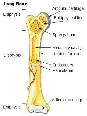
The skeletal system consists of all the bones in are body, cartilage and connective tissue. Bones are actually alive!! Bones, not only support our body, but do so much more. Bones protect internal organs, provide movement, stores mineral reserves and provides a site for blood cell formation. The different types of blood cells, WBC and RBC, are produced in the bone marrow. There are 206 bones in the human body. As I said above bones are alive. They are a solid network of living cells and protein fibers that are surrounded by deposits of calcium salts. A typical long bone, has a shaft and is called the diaphysis. The diaphysis has a medullary cavity which has walls made up of compact bone and is lined with a vascular membrane called the endosteum and is filled with yellow bone marrow which stores fat. The end of a bone is called the epiphysis and this portion of the bone contains spongy bone which has red bone marrow, where blood cells are made. They are also coated with hyaline cartilage, which is called articular cartilage because it occurs at joints and helps reduce friction. All of the bone is covered with a fibrous connective tissue called periosteum, but the ends are not covered with the periosteum. The periosteum actually has four functions (1) shares blood supply, (2) tendons attach to periosteum,(3) periosteum increases diameter as bone grows, (4) it forms callus on the outside, which is if a the bone should break it forms a callus that is very strong! There is also a epiphyseal cartilage on a bone, which is a piece of hyaline cartilage between the epiphysis and diaphysis. It is important because it is responsible for the growth in the length of the bone. There is also a epiphyseal line which shows evidence that the epiphyseal cartilage existed.
Bone is on of the four major connective tissue types. It has a lot of extracellualr matrix. Compact bone is very dense and strong and makes up the majority of the diaphysis of long bones. Compact bone is also composed of tubular units called osteons. Inside the tubular unit are osteocytes which lie in the lacunae. Lacunae are tiny chambers arranged in concentric circles around a central canal. The osteocytes are the cells that live in bone tissue. They maintain the bone matrix and repair the bone if it breaks! The tiny canals are called canaliculi. These small canal through which extensions of osteocytes pass. They run through the bone matrix between the lacuna (hollow space) where the body of the osteocyte is found and the haversian canal where the blood vessels are found. This provides a way for cells to receive nutrients and get rid of wastes. Spongy bone is less dense then compact bone. It is usually found at the end of long bones and contains red bone marrow. It has a lot of thin plates called trabeculae separated by unequal spaces. Cartilage is not as strong as bone, but it is flexible because the matrix is gel-like and contains many collagenous and elastic fibers. Cartilage is avascular, meaning it has no nerves or blood vessels, it gets what it needs from the matrix. There are 3 different types of cartilage. Hyaline cartilage is firm and flexible. Fibrous cartilage is stronger then hyaline because the matrix contains wide rows of thick collagen fibers. Elastic cartilage is flexible and is found in the earlobe and epiglottis in our throat. Fibrous connective tissue are tissue that are closely compact collagenous fibers. It is found in ligaments that connect bone to bone and in tendons where it connect muscle to joints; aka articulations. Besides long bones there are short bones, irregular bones, and flat bones.
Development, growth, remodeling and repair of bone
Bone are made up of different types of cell that help with growth, remodeling and repair. Osteoblasts are bone forming cells. They secrete organic matrix of bone and promote the deposition of calcium salts into the matrix. Next, there are osteocytes and these are mature bone cells made up of osteoblasts. Another type of cell that helps with growth, remodeling, and repair are osteoclasts which are bone absorbing cells. They break done bone and help in depositing calcium and phosphate in the blood. Bone develop by ossification. There are two types of ossification. Intramembranous ossification which bones develop between sheets of fibrous connective tissue. The connective tissue becomes osteoblasts, which I just explained; secret the organic matrix of bone. The secretion is composed of mucopolysaccharides and collagen fibrils. This secretion is added to calcium salts which results in calcification. Through the process of ossification a trabeculae of spongy bone is formed. Spongy bone is on the inside of a bone. The periosteum that is outside the bone creates more osteoblasts to continue with the ossification. The trabeculae fuse together to be come compact bone. The other form of ossification is endochondral ossification and most of our bones are formed this way. This process is where cartilage becomes bone. The cartilage is replaced by the calcified bone matrix that makes these bones capable of bearing weight. There are also steps to the process of endochondral ossification. The first step is the cartilage model. Here chondrocytes lay down hyaline cartilage and is shaped like the future bones or cartilage models. When the cartilage is calcified the chondrocytes die off. Then the bone collar is formed. The osteoblasts secrete the organic bone matrix and the matrix undergoes calcification. This is how the bone collar is formed. This collar covers the diaphysis portion of the bone and will thicken with time. The primary ossification center is where the first bone formation takes place. The medullary cavity and secondary ossification sites are where the spongy bone of the diaphysis is absorbed by osteclasts and that cause the medullary cavity is formed. Lastly is the epiphyseal (growth) plate. Here there is cartilage left between the epiphysis and the diaphysis of a bone. The limb keep increasing in length as long as growth plates are still present. When the epiyseal plate closes, the bone no longer grows in length. There is a very important hormone that plays a key role in the growth of the epiyseal plate and that is the "growth hormone." The growth hormone is released from the anterior pituitary gland. This hormone stimulates growth. Too much of this hormone can cause a person to become gigantism or too little causes dwarfism.

The picture shows a great example of how bones grow! Bones are constantly being broken down by osteoclasts and reformed by osteoblasts in an adult. This is bone remodeling and helps keep bones strong. This process goes on through our life time where new bone is created and old bone is removed. There are two stages that go along with this process and that is absorption and formation. Those two words go along with osteoblasts and osteoclasts because osteoblasts are bone forming cells and the osteoclasts are the bone absorbing cells. The bone recycling also allows the body to regulate the amount of calcium in the blood. Calcium is stored in bones and can be released when it is needed by the body. The parathyroid releases calcium into the blood stream and can control the amount released. There is another hormone that works opposite of calcium and that is calcitonin. Osteoporosis can occur with age, especially in women because estrogen (female hormone) can increase the number of osteoblasts. With the reduction of estrogen the bones of women can be become weaker. Bone remodeling is also why bones can with stand stress. While doing physical activities it enlarges the bone in diameter at the region most affected by an activity. Bone repair is required if someone gets a fracture. This process takes a long time, but the bone will heal. When someone sustains a fracture the first thing that happens is blood vessels are broken inside the bone and this blood forms a hematoma (bloodclot). The repair of the bone starts with the fibrocartilaginous callus. This fills the space between the ends of the broken bone. Next,, the osteoblasts produce the trabeculae of spongy bone and convert fibrocartilage callus to a bony callus. This joins the broken pieces together. Remodeling is the last step. Osteoblasts build new compact bone and osteoclast absorb the spongy bone and create a new medullary cavity. The use of casts also helps in setting a bone while it is repairing. The cast makes sure that the limb has no movement so it can heal. There are different types of fracture. Below is pictures of the different kinds.

Bones of the axial skeleton consists of the skull, hyoid bone, vertebral column, and the rib cage. The are many different bone the make up the skull. The skull also referred to as the cranium protects the brain. The major bones of the cranium have the same name as the lobes of the brain. They are the frontal bone(forehead), the parietal bone (sides), occipital bone (back of head), temporal bone (where our ears are), sphenoid bone (floor of cranium), and the ethmoid bone (in front of sphenoid). To understand this better it helps to have a picture.............

There are also bone of the face. The mandible(our jaw), zygomatic bones (cheekbones), and the vomer (nose area). All of these bones help protect the brain and give our face its shape. The hyoid bone, which is not part of the skull, can be found in our throat area. The is the only bone that does not articulate with another bone. It is attached to the temporal bone by ligaments and to the larynx by a membrane. The hyoid bone anchors the tongue and serves as the site for the attachment of muscles associated with swallowing. if this bone happens to get broken, it is an indication of strangulation. The vertebral column is responsible for housing the spinal cord and the bone of the column protect the cord. The vertebral column consists of 5 different bone. The cervical bones are in the neck area and there are 7 of them with the first bone being the atlas (holds the head) and the 2nd bone is the axis (helps with turning of the head). The thoracic bones (12bones) which have long spinous processes articulate with the ribs. The lumbar bones (5 bones) have a large body and help with support of the body. The sacral bones (5 bones) are fused with the sacrum. The last bone of the spinal column is the coccyx (tailbone) and is composed of four fused vertebrae.

Not shown in this picture is the intervertebral disc which are composed of fibrocartilage that helps cushion the bones. It also prevents grinding of bones together and absorbs shock like when someone is jumping or running. The rib cage helps protect all the organs underneath it. It is composed of thoracic vertebrae, ribs, cartilage, and the sternum. There are 12 pairs of ribs and these articulate with the thoracic bones in the spinal column. The last set of ribs don not attach they are called floating ribs. The sternum is a flat bone that lies in the middle of the chest and where the "true ribs" are attached. The sternum is made up of 3 bones. The manubrium (the handle), the body (the blade), and xiphoid process (the pointy part at the end). The manubrium articulates with the clavicle of the appendicular skeleton and the first pair of ribs.
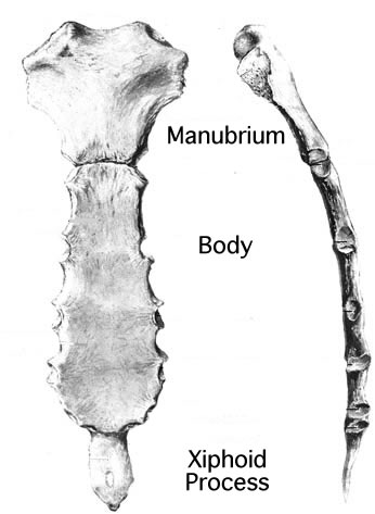
www.yorku.ca/earmstro/journey/images/sternum
Bones of the appendicular skeleton consists of the pectoral and pelvic girdles and their attached limbs. With the pectoral girdle, left and right sides, there is the scapula (shoulder blade), and the clavicle (collarbone). It also consist of the arm bones. The humerus (long bone in arm), radius and ulnar (forearm bones), carpal and metacarpals (bones in hands) and the phalanges (also bones in fingers).

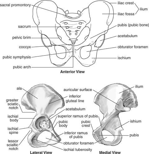
The muscles of the arm and chest attach to the coracoid process of he scapula. The glenoid cavity of the scapula articulates with the head of the humerus. As seen in this picture there are many bones in the hands, each with their own special name besides carpals. The pelvic girdle and lower limb consists of the pelvis (hip girdle) and the bones of the leg. The pelvis is composed of the sacrum and the coccyx which you can see in the picture above in the vertebral column. There are three parts to the pelvic; the ilium, the ischium and the pubis. The socket that the hip bone fits into is called the acetabulum. We sit on the ischium. The pubis symphysis is where the two pubic bones are joined together by a fibrocartilaginous joint. The leg consists of the femur (long bone), tibia & fibula (calf bones), tarsals and metatarsals (make up the foot), and phalanges (foot bones).
 media.wiley.com
On the leg there is also the knee bone which protrudes from your leg and is called the patella. The foot also has other bones besides the tarsals. The ankle bone (talus), and heel bone (calcaneus). These two bones support the weight of the body. All the bones in our body serve a purpose, not only to support our body, but protect our organs, allows movement, and store important minerals. Bones are joined at the joints, which are cartilaginous or synovial. This is what allows our arm and legs to move freely, but there are different types of movement. Our arms can rotate, but our legs can't. Here is a picture of movements that our body allows.....

These are not the only movements that our body allows. There are different movements for hands and feet too. The skeletal system works very closely with the muscular system.
THE MUSCULAR SYSTEM
A. types of muscles & function
B. skeletal muscle of the body
C. muscle fibers and how they slide
D. motor units
E. energy for muscle contraction
F. Fast and Slow twitch muscle fibers
G. common muscular conditions
H. Muscular diseases
There are three types of muscle in the human body; skeletal, cardiac, and smooth. Skeletal muscle fibers are tubular, multinucleated and striated. They attach to bone. Skeletal muscle is voluntary because we move a part of the body when we want to. Skeletal muscle is responsible for thermoregulation, which maintains our body temperature for our nerves to work properly. It is also responsible for movement, maintains posture, and stabilizing joints. The skeletal muscle assists movement of cardiovascular and lymphatic vessels through pressure gradients. Cardiac muscle is only in the heart (walls of the heart) and it is responsible for pressure gradients and excitatory fibers. It has a single nucleus, striated, tubular, and branched which allows fibers to interlock at intercalated disks. Cardiac muscle is involuntary because we don't have control of its contractions. Smooth muscle is also has a single nucleus and their cells are in a parallel line, forming sheets. It is found within the wall of internal hollow organs. It is involuntary just like cardiac muscle. Here are pictures of the three types of muscles..........


Skeletal muscle is made up of fibers called fascicles. Within the fascicles are fibers that are covered with connective tissue. Skeletal muscles vary in size, shape, and arrangement of fibers. The muscles are covered with fascia which is a form of connective tissue that extends to become a tendon. Skeletal muscles work in pairs. There is an insertion which is the muscle is on the bone that moves and the origin which is the muscle is on a stationary bone. Muscles are responsible for contraction and when the muscle contracts it shorten. There is a primary mover and a synergist (assist the prime mover) when a muscle contracts. How do muscle fibers slide? There are quite a few things that go into the sliding of muscle fiber. Each muscle fiber contains a large number of myofibrils that are composed of sarcomeres. The sarcomere is the basic unit of the contraction and the subunits of myofibrils. The cell membrane is called the sarcolemma and the endoplasmic reticulum is called the sarcoplasmic reticulum (where calcium is stored). The sarcolemma forms the transverse tubules that penetrate into the cell so that they come into contact with the sarcoplasmic reticulum. The myofibrils are bundles of overlapping myosin and actin filaments. There are four proteins of a sarcomere. Myosin is the thick filament protein and actin is the thin filament protein. The next two are troponin and tropomysin and these regulate myosin and actin interaction. There are also two supporting proteins that participate in a muscle contraction; (1) titin is elastic and helps return a stretched sarcomere to resting length;(2) nebulin and it serves to align the actin filaments. Also, there is an organization of a sarcomere; the Zdisks serve as attachment sites for actin filaments; I band (actin only) region occupied only by thin filaments; A band is where overlapping of myosin and actin occurs and encompasses the thick filaments; H Zone (myosin only) ; M Line attachment site for thick filaments
CONTRACTION OF A MUSCLE
Muscle fibers are stimulated to contract by motor neurons whose axons are in nerves. The axon of one neuron can stimulate little or a lot of muscle fibers of a muscle because each axon has different branches. A small gap called a synaptic cleft separates the axon terminal from the sarcolemma. This is known as the neuromuscular junction. Ach (acetylcholine) is a neurotransmitter that is release though the synaptic cleft and binds to receptors in the sarcolemma. The sarcolemma generates impulses that spread over the sarcolemma and down the T tubules to the sarcoplasmic reticulum. Contraction begins when Ca2+ (calcium released from the sacroplasmic reticulum) binds to troponin causing tropomyosin to be pulled towards the groove of actin filaments. This exposes the myosin binding site on actin so that myosin can bind to complete the power stroke. Myosin has a ATPase site so ATP can attach. The myosin head tightly bound to actin molecule. Myosin has two functions end tail and head. ATP binds with the nucleotide binding site inducing change in the myosin head so that it no longer binds with actin. The nucleotide binding site on myosin closes around the ATP and hydrolysis it to ADP & P, both remain bound to myosin. The energy released from ATP rotates the free myosin molecule and it binds weakly to a new actin molecule one or two positions away. The power stroke is initiated when a phosphate is released from the myosin binding site. As myosin moves it pushes the attached actin filament toward the center of the sarcomere. The myosin releases ADP and is now tightly bound to the actin. The cycle will repeat with the binding of another ATP. Contraction is regulated by troponin and tropomyosin. In the relaxed state tropomyosin blocks the myosin binding site on actin.


MOTOR UNITS
A motor unit consists of all the muscle fiber innervated by one motor neuron. All fibers of a motor unit respond to a stimulus. The number of muscle fibers per unit ranges from 4 to several 100. Fibers in a unit are spread throughout the entire muscle. The greater the number of fibers contracting the greater the total muscle tension. A single action potential in a muscle fiber produces a brief contraction called a "twitch." With more and more contraction, the more motor units are needed. A muscle twitch is divided into three stages; the latent period which is the time between a stimulus and initiation of contraction; the contraction period is when the muscle shortens; and the relaxing period is when the muscle returns to its normal length. If a motor unit receives multiple stimuli it can respond without relaxing. If a muscle fiber is stimulated so rapidly that it does not have an opportunity to relax at all between stimuli a maximal sustained contraction known as a tetanus occurs. A tetanus continues until the muscle fatigues.
ENERGY FOR MUSCLE CONTRACTION
Resting muscle stores energy from ATP in high energy phosphate bonds of phosphocreatine. Working muscle transfers energy from phosphocreatine back to ATP. Creatine phosphate is the first energy storehouse tapped into at the onset of contractile activity. Like ATP creatine phosphate contains a high energy phosphate group which can be donated to ADP to form ATP. There are other energy sources for muscle contraction. There are two that are in our blood; glucose and plasma fatty acids. Both of these are delivered through blood circulation. People that exercise to lose weight use up the plasma fatty acids which are adipose tissue (fat) that makes us look fat. When the diet is restricted it uses up the adipose tissue that is stored. Fermentation happens when someone is exercising for a long period of time and feeling a burning sensation, that is the lactate building up. Fermentation also makes the muscle fatigued. While working out most people breath heavy and this is called oxygen debt, which is the bodies way of completing the metabolism of lactate and restore the cells to their original energy state. Fermentation is an anaerobic process.
FAST AND SLOW TWITCH MUSCLE FIBERS
With the fast twitch muscle fibers they are dependent on anaerobic pathways, it does not need oxygen to make ATP. The muscle contracts rapidly and tires easily. Common in upper limbs and less endurance. The slow twitch are aerobic and have thin fibers. the slow twitch fibers have many mitochondria and myoglobin giving it a dark color. Myoglobin is similar to hemoglobin and can store small amounts of oxygen, but more importantly it increases the rate of oxygen transfer from the blood to the muscle fibers.
COMMON MUSCULAR CONDITIONS
I believe that many, if not all people, have suffered from one of these muscular conditions. Spasms are a sudden involuntary contraction of a muscle. They don't last long, but you know they are there. Cramps are strong painful spasms. They can be caused by strenuous exercise or not enough potassium in your diet. A strain is caused by over stretching a muscle. Lastly, a strain is when you twist your ankle and it swells.
MUSCULAR DISEASES
Muscular dystrophy is a progressive disease that affects the muscles. The muscles fibers die and is replaced by fat and connective tissue. Another disease is Duchenne muscular dystrophy and is most common because it is inherited by a gene carried by the mother. A protein called dystrophin causes the condition. When the protein is absent calcium leaks into the cell and activates an enzyme to dissolve muscle fibers.
The skeletal and muscular systems work very closely. They rely on each other. The skeletal system protect our internal organs and the muscular system pads bones.


2 comments:
Post a Comment